
Vitrectomy Surgery
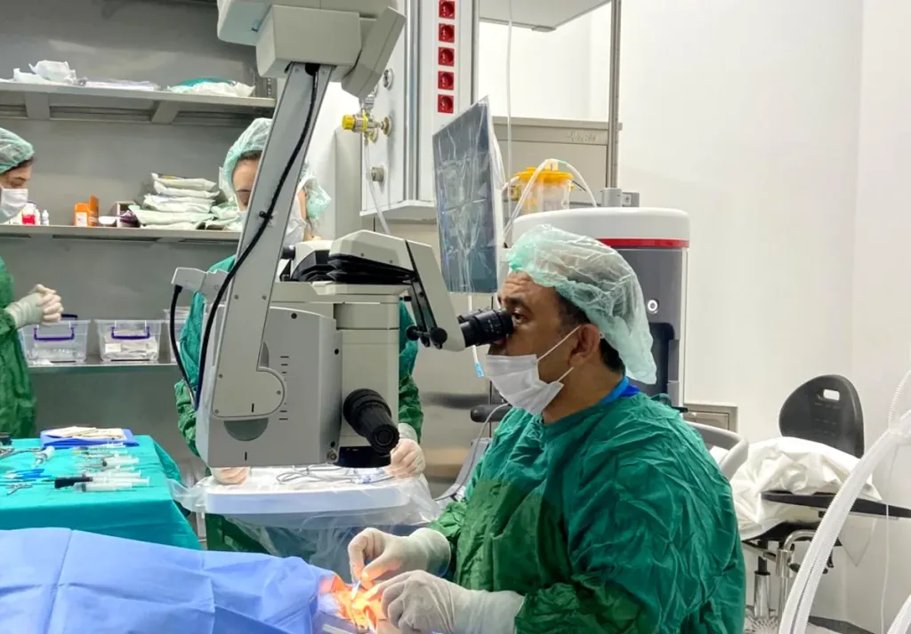
By Dr. Nejmi Öztürk, ophthalmologist and eye surgeon, at HG Hospital in Kahramanmaraş, Türkiye — surgery for retinal detachment, diabetic bleeding, macular hole and epiretinal membrane
- What vitrectomy is and which retinal diseases it is done for
- Steps of the operation, type of anesthesia and duration
- Gas and silicone oil tamponade: face-down positioning and the rule about air travel
- Recovery timeline, risks and insurance information
What is vitrectomy?
Vitrectomy is the removal of the clear gel that fills the eye (the vitreous) through incisions smaller than a millimeter in the white of the eye, and the repair of the problem in the retina (the seeing layer on the back wall of the eye). The vitreous gel is not needed for vision; the space it leaves fills with the fluid the eye produces itself. At the end of the operation, gas or silicone oil is placed inside the eye if needed to hold the retina in place.
The operation is done under a microscope with fine instruments that enter the eye. Today small-diameter systems known as 23, 25 and 27 gauge are used; the incisions usually close without stitches. For an overview of retinal diseases, see the retinal diseases page.
When is vitrectomy done?
Vitrectomy is done when the retina has detached, when there is bleeding inside the eye, when a hole or membrane has formed in the macula, and when there is an infection or a foreign body inside the eye.
- Retinal detachment (the retina separating from its place): Fluid passing through a tear in the retina lifts it off the wall; with vitrectomy the retina is laid back in place and the tear is sealed with laser.
- Diabetic vitreous hemorrhage: Bleeding from the new vessels caused by diabetes fills the inside of the eye and blocks vision; the blood is cleared and the retina is treated with laser. If a tractional detachment has developed, the membranes are removed.
- Macular hole: A small hole that forms at the center of vision; the gel and the inner membrane are peeled so that the hole can close, and a gas tamponade is used.
- Epiretinal membrane (a membrane in front of the macula): A thin membrane that forms on the surface of the retina wrinkles it and causes distorted vision; the membrane is peeled off.
- Infection inside the eye (endophthalmitis) and foreign bodies: The infected gel is cleared and medicine is given; in injuries, the foreign body inside the eye is removed.
- Other: Lens fragments that have dropped into the eye, a dislocated intraocular lens, bleeding under the macula.
Who is it not suitable for?
Vitrectomy is not suitable for eyes that have no remaining potential for vision or for patients whose general condition cannot tolerate anesthesia; it is also generally not recommended for ordinary floaters.
- Eyes in which the optic nerve or retina is permanently damaged and no gain in vision is expected from surgery
- Patients with uncontrolled blood sugar, serious heart or lung disease or a bleeding disorder: medical preparation is needed beforehand
- People with an active infection on the outside of the eye (eyelid and conjunctiva): surgery is not started until the infection has been treated
- People who have nothing more than bothersome floaters: the balance of risk and benefit is usually not in favor of surgery
Which examinations and tests are done before the operation?
Before the operation, a dilated retinal examination is done and the condition of the retina is documented with OCT and, if necessary, an eye ultrasound; a medical assessment and blood tests are requested for anesthesia.
- Retinal examination: The location of the tears, the extent of the detachment and whether the macula is involved are determined.
- OCT: A macular hole, epiretinal membrane and edema are seen in detail; the images are recorded for comparison after surgery.
- Eye ultrasound: When the retina cannot be seen because of dense bleeding, it shows whether the retina is in place.
- Eye pressure and anterior segment examination: If there is a cataract, cataract surgery may be planned in the same session.
- Anesthesia assessment: Blood tests, ECG and adjustment of blood-thinning medicines.
How is vitrectomy surgery performed?
Vitrectomy is done in the operating room, mostly under local anesthesia given by an injection around the eye together with light sedation, and in some patients under general anesthesia; depending on the complexity of the disease it takes between 30 minutes and 2 hours.
- Anesthesia and preparation: The area around the eye is numbed; the eye and eyelids are cleaned with antiseptic and a sterile drape is placed. General anesthesia is preferred for children, for operations that will take a long time and for patients who cannot lie on their back.
- Micro-incisions: Three small entry ports are made in the white of the eye, a few millimeters behind the edge of the cornea. Through one, fluid is supplied to the eye; through another, light; through the third, the working instrument.
- Removal of the gel: The vitreous gel is cleared with a fine instrument that cuts and aspirates thousands of times per second; blood and cloudiness are removed at this stage.
- Repair of the retina: Tears are located and surrounded with laser; fluid under the retina is drained; in macular hole and epiretinal membrane surgery the membrane on the retinal surface is stained with a special dye and peeled; in diabetic patients the membranes causing traction are removed.
- Tamponade: If the retina needs to be held in place, air, gas or silicone oil is placed inside the eye. Which one is used is chosen according to the disease.
- Closure: The small incisions usually close by themselves; if necessary a dissolving stitch is placed. The eye is covered and a protective shield is fitted.
Under local anesthesia you are awake; pain is not expected, while a feeling of pressure and flashes of light are normal. If there is any discomfort, the anesthesia team gives additional medicine. Patients are usually discharged the same day or the next morning.
What are gas and silicone oil tamponade, and why is positioning important?
A tamponade is a gas bubble or silicone oil that holds the retina in place by pressing it against the wall while it heals. Because gas rises, the head is positioned so that the area of the tear is uppermost; for a macular hole this means face-down positioning. The duration and type of positioning are decided by your doctor and usually cover most of the day, for anywhere from a few days to 1–2 weeks.
| Tamponade | Time in the eye | Vision | Flying and altitude | Further surgery |
|---|---|---|---|---|
| Air | A few days | Blurred until the bubble is absorbed | Not allowed (while the bubble is present) | Not needed |
| Short-acting gas | About 2–3 weeks | Clears from the bottom up as the bubble shrinks | Not allowed until the gas is completely absorbed | Not needed |
| Long-acting gas | About 6–8 weeks | Blurred for weeks | Not allowed until the gas is completely absorbed | Not needed |
| Silicone oil | Months (planned) | Blurred; glasses prescription changes | Allowed | The oil is removed in a second operation |
Vitrectomy step by step
Removing the vitreous, repairing the retina and supporting the inside of the eye with a tamponade.
- Identifying the problemSurgery comes into question when the retina has detached, or when there is bleeding or traction within the vitreous. The extent is determined with examination and imaging.
- Entry through three small portsThree small ports made in the white of the eye are used: one for fluid balance, one for light and one for the cutting-suction instrument. Stitches are not needed in most cases.
- Support with a tamponadeAfter the vitreous is removed, the eye is filled with gas or silicone oil; this tamponade holds the retina in place. Gas is absorbed on its own, while silicone oil is removed in a second procedure.
What is recovery like after vitrectomy?
After vitrectomy, vision is blurred for weeks until the tamponade is absorbed and the retina has settled; the final level of vision is reached in 3–6 months depending on the disease, and in macular diseases it can take up to a year.
- Day 1: The shield is removed and the eye and retina are checked; eye pressure is measured. Stinging, redness and swelling of the eyelid are normal. Antibiotic and cortisone drops are started; the positioning rule is followed.
- Week 1: Pain decreases; vision is very low in an eye with gas and blurred in an eye with silicone oil. Heavy lifting, bending, rubbing the eye and swimming pools are not allowed. At the check-up the retina and the incisions are assessed.
- Month 1: As short-acting gas is absorbed, vision clears from the bottom upwards; the drops are reduced. Return to desk work is usually after 2–4 weeks; the flying ban continues until the gas has gone.
- Months 3–6: Vision settles; new glasses are prescribed in this period. If silicone oil was used, the time for its removal is planned; the development of cataract is monitored.
For patients coming from Elbistan, Afşin, Göksun and the surrounding districts, the day 1 check-up is planned together with the operation; if you will be returning over high mountain roads with gas in the eye, your doctor must be informed in advance.
What are the risks and limitations?
Vitrectomy is a major operation inside the eye; its purpose is to save the retina, but it cannot bring vision back in every patient, and repeat surgery may be needed.
- Development of cataract: In most patients who still have their natural lens, a cataract progresses within 1–2 years; cataract surgery may be needed.
- Rise in eye pressure: May occur because of the gas, the silicone oil or the cortisone drops; it is controlled with drops.
- Repeat detachment: If healing tissue forms on the surface of the retina (proliferative vitreoretinopathy), the retina can detach again; further surgery is needed.
- Infection (endophthalmitis): Rare but sight-threatening; seek care immediately if pain, redness and loss of vision increase.
- Bleeding, a new retinal tear, failure of the macular hole to close, recurrence of the membrane: Seen with a frequency that varies according to the disease.
- Problems related to silicone oil: Oil passing into the front of the eye, corneal damage, raised eye pressure.
- Anesthesia risks and, rarely, loss of vision.
Limitations: In detachments where the macula has been detached for a long time, and in advanced diabetic damage, the gain in vision may be limited even if the retina is reattached. The visual result depends on the degree and duration of the damage before the operation.
What are the alternatives to vitrectomy?
Depending on the disease, smaller procedures can be used instead of, or before, vitrectomy; the choice depends on the location of the tear, the extent of the detachment and the patient’s condition.
| Situation | Alternative | When it is preferred | Details |
|---|---|---|---|
| Retinal tear (no detachment) | Retinal laser or freezing (cryotherapy) | When the tear is caught early; done as an outpatient | Retinal diseases |
| Limited retinal detachment | Gas injection (pneumatic retinopexy) or scleral buckling | When the tear is at the top and single; in young patients who still have their natural lens | This page |
| Diabetic bleeding | Waiting, with anti-VEGF injection and retinal laser | When the bleeding is slight and the retina can be seen | Diabetic retinopathy |
| Mild epiretinal membrane | Observation | When vision is good and symptoms are few | This page |
Vitrectomy in Kahramanmaraş
Vitrectomy surgery is performed at HG Hospital in Kahramanmaraş, Türkiye, by ophthalmologist Dr. Nejmi Öztürk. The pre-operative retinal examination, OCT and the necessary tests are done at the same hospital; in emergencies such as retinal detachment, the operation is planned without delay.
HG Hospital is a private hospital contracted with the Turkish social security institution (SGK); vitrectomy is within SGK coverage, and patients with valid SGK coverage may use it, although a private-hospital co-payment may apply. Patients without SGK coverage, such as international patients and visitors from abroad, pay privately or through their private or travel health insurance; the hospital checks coverage before treatment. Fee information is given by the hospital after the examination, and insurance coverage is clarified at the consultation.
Patients from Elbistan, Afşin, Göksun, Ekinözü and Nurhak come to HG Hospital in Kahramanmaraş city center for the examination and the operation; Dr. Öztürk does not see patients in Elbistan. For the day of surgery, the day 1 check-up and accommodation planning, see the Elbistan and surrounding area page.
Dr. Nejmi Öztürk sees patients at HG Hospital in Kahramanmaraş, Türkiye. For patients travelling from other districts or from abroad, examination, tests and treatment planning can be arranged for the same day.
Hospital switchboard (within Türkiye): 444 46 46 · E-mail: info@nejmiozturk.com · HG Hospital, Üngüt Mevkii, Prof. Dr. Necmettin Erbakan Blv. No:209, Onikişubat / Kahramanmaraş, Türkiye
Frequently asked questions
How many hours does vitrectomy surgery take?
Clearing a simple hemorrhage takes 30–45 minutes, epiretinal membrane and macular hole surgery about 1 hour, and extensive detachments and advanced diabetic cases 1.5–2 hours. The time can be longer depending on what is found during the operation.
Will there be pain during the operation, and will I be asleep?
Most patients have local anesthesia by an injection around the eye together with light sedation; you are awake, pain is not expected, and you feel pressure and see light. General anesthesia is preferred for children, for long operations and for patients who cannot lie on their back. The type of anesthesia is decided by the surgeon and the anesthesia team together.
How long does face-down positioning last, and how can it be made easier?
The positioning period varies from a few days to 1–2 weeks depending on the disease and the gas used; your doctor tells you how many hours a day and in which direction to position. Face-down pillows, sitting at a table with forehead support and short breaks every hour make the process easier. Keeping to the positioning is important for the hole to close and the retina to stay attached.
Why can’t I fly while there is gas in the eye?
Cabin pressure drops in an aircraft; the gas inside the eye expands and raises the eye pressure suddenly and dangerously. This can permanently damage the optic nerve. The ban lasts until the gas has been completely absorbed, which is confirmed at a check-up with your doctor; the same rule applies to high mountain roads.
When is the silicone oil removed?
Silicone oil is usually removed in a second operation within 3–6 months, once the retina has settled; in some complex cases it may be left for longer. As long as the oil is in the eye, vision is blurred and the glasses prescription is different. The timing of removal is decided at the retinal check-ups.
When does vision improve after vitrectomy?
In an eye with gas, vision clears over 2–8 weeks as the bubble is absorbed; in an eye with silicone oil it remains blurred until the oil is removed. After retinal detachment the final vision settles in 3–6 months; after macular hole and epiretinal membrane surgery it can take up to a year. The level of vision that can be reached depends on the degree of damage before the operation.
Does a cataract develop after vitrectomy?
In the large majority of patients who still have their natural lens, a cataract progresses within 1–2 years after vitrectomy. This is expected and is treated with cataract surgery. In some patients vitrectomy and cataract surgery are planned in the same session.
How urgent is retinal detachment surgery?
If the center of vision (the macula) has not yet detached, the operation is done within days, usually within the first 24–48 hours; if the macula has detached, it is still planned without delay. The longer the wait, the smaller the gain in vision. If you suddenly feel a curtain coming down over your vision, see an eye doctor the same day.
Is vitrectomy covered by SGK, and how much does it cost?
Vitrectomy is within SGK coverage for insured patients; a private-hospital co-payment may apply. Fee information is given by the hospital after the examination; insurance coverage is clarified at the consultation. Patients without SGK coverage pay privately or through their private or travel health insurance.
I am coming from Elbistan; what about travel and check-ups after the operation?
Dr. Öztürk sees his patients only at HG Hospital in Kahramanmaraş city center; Elbistan is about 148 km, or 2 hours, away. For the day 1 check-up, you plan either to stay in the city or to come back the next day. While there is gas in the eye, ask your doctor about the high passes on the way home and keep to the positioning rule as much as possible during the journey.
Sources
- Turkish Ophthalmological Society — todnet.org
- American Academy of Ophthalmology (AAO) — aao.org
- National Eye Institute (NEI) — nei.nih.gov
Related pages
This page is for information only; diagnosis and treatment decisions are made after an examination. The results of any surgical or interventional procedure may vary from person to person. You are advised to obtain detailed advice from your physician before any procedure.

Ophthalmologist and eye surgeon. Graduate of Erciyes University Faculty of Medicine; completed his residency at Ankara Atatürk Training and Research Hospital. With 21 years of experience in cataract and refractive surgery, strabismus, glaucoma, retina and oculoplastic surgery, he sees patients at HG Hospital in Kahramanmaraş, Türkiye. Biography and training
Dr. Nejmi Öztürk
Ophthalmologist and eye surgeon
HG Hospital, Üngüt Mevkii, Prof. Dr. Necmettin Erbakan Blv. No:209, Onikişubat / Kahramanmaraş
+90 506 377 47 68WhatsAppAppointment formHospital switchboard (within Türkiye): 444 46 46 · info@nejmiozturk.com
- Private patients and private health insurance are welcome; patients with SGK coverage may use it (conditions are confirmed by the hospital)
- Same-day examination and test planning for patients travelling from other districts or from abroad
- Getting here and patient information
- Patients from Elbistan and the region