
ICL (Phakic Intraocular Lens) Surgery
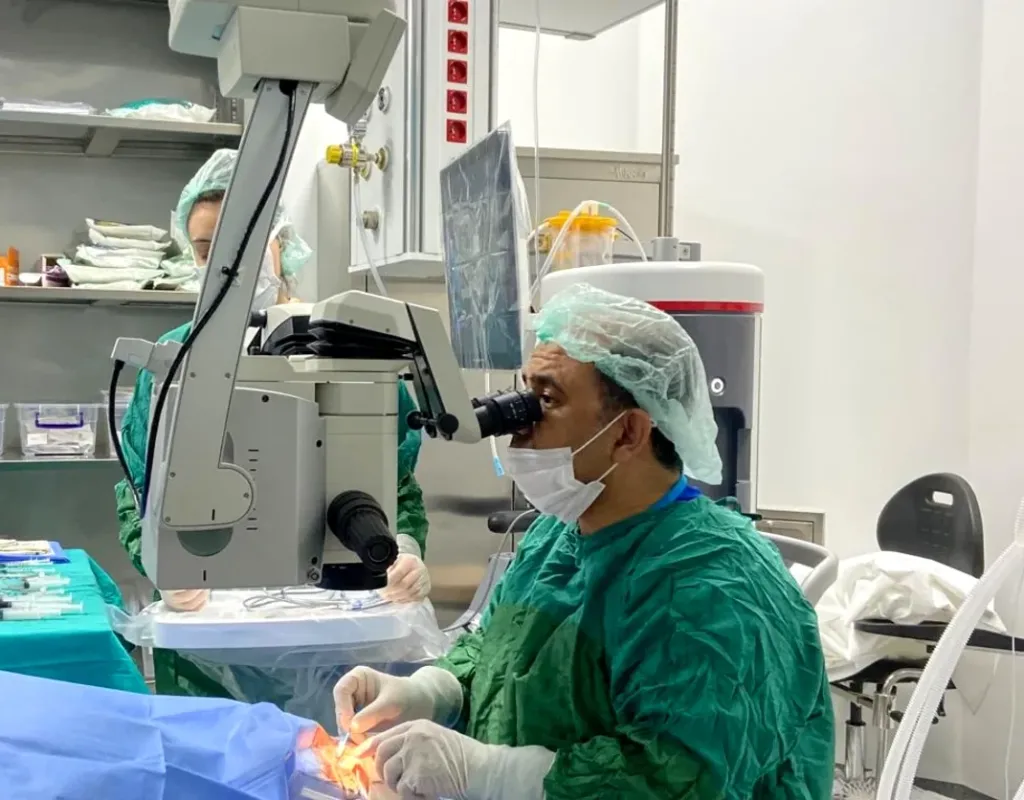
Objective information about the phakic intraocular lens — suitability is assessed with the examination and measurements of Dr. Nejmi Öztürk, ophthalmologist and eye surgeon, at HG Hospital in Kahramanmaraş, Türkiye
- ICL is a thin, flexible lens placed between the iris (the colored part of the eye) and the natural lens, which is left in place; your own lens is not removed.
- It is considered between the ages of 21 and 45 for high myopia, thin corneas and dry eye, when laser eye surgery is not suitable.
- The lens can be removed if necessary; however, yearly check-ups for life are needed for cataract, eye pressure and endothelial cell loss.
- It is generally not covered by SGK; suitability is assessed with measurements such as anterior chamber depth and endothelial cell count.
What is ICL?
ICL (phakic intraocular lens) is a thin, flexible lens placed in the space between the iris and the lens, while the eye’s natural lens is left in place. The word “phakic” means that your own lens is preserved; in this respect ICL differs fundamentally from cataract surgery and refractive lens exchange, in which the lens is removed. The lens corrects the prescription much as a contact lens does; the difference is that it stays permanently inside the eye and needs no daily care.
ICL does not touch the cornea (the clear front layer of the eye) and removes no tissue; it is therefore an option for eyes in which laser is not suitable. The natural lens keeps its ability to change focus. The lens can be removed if necessary; however, being “reversible” does not mean that the eye will return to its original state in every situation.
Who is ICL suitable for?
ICL is suitable for patients aged 21–45 with moderate-to-high myopia (nearsightedness, blurred distance vision) whose prescription has not changed for at least one year and who are not suitable for laser. The main criteria are:
- High myopia (roughly -6 to -18 diopters); astigmatism can also be corrected with a toric ICL
- Corneal thickness or structure found unsuitable for laser
- Marked dry eye (eyes in which laser is not advised because dryness may increase after laser)
- An anterior chamber depth (the space between the cornea and the lens) sufficient for the lens
- An endothelial cell count (the cell layer on the inner surface of the cornea) adequate for the person’s age
- No cataract, glaucoma or active retinal disease
Who is it not suitable for?
ICL is not suitable for eyes with a shallow anterior chamber or a low endothelial cell count, because the lens then sits too close to the cornea and can accelerate cell loss. It is also not recommended in the following situations:
- A cataract that has begun, or clouding of the lens
- Glaucoma (raised eye pressure), a narrow-angle eye structure or persistently high pressure inside the eye
- A history of uveitis (inflammation inside the eye)
- People under 21 whose prescription has not yet settled; over 45, lens exchange is usually considered instead
- Pregnancy and breastfeeding (the prescription can change temporarily)
- An untreated retinal tear or thinning; this is treated with retinal laser first
In patients who do not meet these criteria, laser eye surgery is considered if the cornea is suitable, and lens exchange options for those over 45.
Which examinations and tests are done before surgery?
Before ICL, the power and the size of the lens are calculated; the measurements are therefore more detailed than for a laser examination, and choosing the correct lens size is the foundation of the result.
- Prescription measurement: The true prescription is determined with the pupil dilated by drops; the power of the lens is calculated from this.
- Anterior chamber depth: Measured with anterior segment OCT or optical biometry; the space the lens will fit into must be sufficient.
- Corneal diameter and sulcus measurement: The diameter of the cornea and, if needed, the space behind the iris are measured with ultrasound biomicroscopy; the length of the lens is chosen accordingly.
- Endothelial cell count: The number of cells on the inner surface of the cornea is measured with a specular microscope; this is the baseline value for follow-up.
- Corneal topography: The axis of the astigmatism is determined; for a toric ICL the placement angle is calculated.
- Eye pressure and angle examination: The pressure inside the eye and the angle structure at the root of the iris are assessed.
- Dilated retinal examination: In high myopia the periphery of the retina is examined for tears and thinning.
If you wear contact lenses, you will be asked to leave soft lenses out for at least one week before the measurements, and rigid lenses for longer. With some lens designs a small laser opening in the iris (iridotomy) may be needed beforehand; with newer designs that have a central hole it is usually not necessary.
How is ICL surgery performed?
ICL surgery is done with anesthetic drops through an incision of about 3 mm, takes 15–30 minutes per eye, and you go home the same day. The steps are as follows:
- Drops that dilate the pupil and numb the eye are applied; the area around the eye is cleaned.
- An incision of about 3 mm is made at the edge of the cornea; the inside of the eye is filled with a protective gel.
- The folded lens is delivered into the eye through a fine injector and unfolds slowly.
- The four corners of the lens are positioned in the groove behind the iris (the sulcus); a toric ICL is rotated to the calculated axis.
- The protective gel is rinsed out, and a drop that constricts the pupil is given. The incision seals by itself; stitches are usually not needed.
During the procedure you feel light, mild pressure and touch. Eye pressure is measured a few hours after surgery; a temporary rise in pressure can occur in the first hours. The two eyes can be operated on the same day or a few days apart; because you will not be able to drive after surgery, you need to come with a companion.
ICL surgery step by step
Placement of the implantable lens behind the iris while the natural lens stays in place, shown schematically on a cross-section of the front of the eye.
- Small incisionAn incision of about 3 mm is made at the edge of the cornea. The natural lens is not touched; the eye keeps its own lens.
- Inserting the lensThe soft lens is passed through this incision in folded form and unfolds inside the eye.
- Final positionThe lens sits between the iris and the natural lens. It is not visible or felt from outside, and it can be removed if needed.
What is recovery like?
In most patients vision clears noticeably within 1–2 days, because there is no surface wound on the cornea that needs to heal. Check-ups are on the day after surgery, at week 1, at month 1 and at month 3–6; after that a yearly check-up is needed for life.
- Day 1: Eye pressure, the position of the lens and the distance between the lens and the natural lens are measured. Antibiotic and anti-inflammatory drops are started; mild haziness and light sensitivity are normal.
- Week 1: Do not rub your eyes, keep water out of your eyes and do not lift heavy loads. Screens and reading are allowed.
- Month 1: The drops are tapered and stopped; wait one month before swimming pools, the sea and contact sports. Any remaining prescription is measured.
- Yearly check-up: The endothelial cell count, eye pressure, the lens distance and the clarity of the natural lens are assessed every year.
If you are coming from Elbistan, Afşin, Göksun, Ekinözü or Nurhak, the day of surgery and the day 1 check-up are planned together; the later check-ups are arranged according to your travel situation.
What are the risks and limitations of ICL?
Because ICL is a structure placed inside the eye, its risks differ from those of laser, and some of them can be noticed only at the yearly check-ups; keeping to the follow-up is therefore part of the surgery.
- Cataract development: If the lens sits too close to the natural lens, or over the years, clouding can develop on the front surface of the natural lens; the ICL is removed and cataract surgery is performed.
- Rise in eye pressure: In the first days the pressure can rise because of residual gel or the drops, and in the long term because the lens affects the flow of fluid; drops or an additional procedure may be needed.
- Endothelial cell loss: The cells on the inner surface of the cornea can decrease over time; if the count falls below a certain level, the lens is removed. This is why the yearly count is mandatory.
- Wrong lens size: If the lens sits too high or too low, an exchange may be needed.
- Halos and glare: People with large pupils may see rings around lights at night; this usually decreases.
- Rotation of a toric lens: If the astigmatism correction is lost, the lens is repositioned.
- Infection and retinal detachment: Rare; in high myopia the risk of retinal detachment is higher regardless of surgery.
As a limitation, ICL does not correct difficulty with near vision (presbyopia); reading glasses will be needed after the age of 45. Small remaining prescriptions are corrected with glasses or, if the cornea is suitable, with laser.
ICL or laser? A comparison
The basic difference between ICL and laser is that laser reshapes the cornea, whereas ICL adds a lens inside the eye without touching the cornea.
| Feature | ICL (phakic lens) | Laser (SMILE, LASIK, PRK) | Refractive lens exchange |
|---|---|---|---|
| What is done? | A lens is added in front of your own lens | The cornea is reshaped with laser | The natural lens is replaced with an artificial lens |
| Suitable prescription | Moderate–high myopia, astigmatism | Low–moderate myopia, hyperopia, astigmatism | High hyperopia, over 45 |
| Thin cornea | Suitable | Limiting | Suitable |
| Dry eye | No effect | May increase it | No effect |
| Reversibility | The lens can be removed | Permanent | Permanent |
| Long-term follow-up | Yearly check-up mandatory | Routine examination | Routine examination |
| SGK coverage | Generally not covered | Generally not covered | Partly, if a cataract is present |
For details of the laser methods, see the SMILE and PRK pages; for eyes with suspected keratoconus, see the keratoconus treatment page.
Assessment and planning in Kahramanmaraş
Your suitability for ICL is assessed with the examination and measurements at HG Hospital in Kahramanmaraş, Türkiye; the method that suits you is planned together. The examination is done by Dr. Nejmi Öztürk, an ophthalmologist working in cataract and refractive surgery; depending on the results, the options of ICL, laser or lens exchange are discussed with you.
Because ICL is a procedure to reduce the need for glasses, it is generally not covered by SGK. HG Hospital is a private hospital contracted with the Turkish social security institution (SGK); patients with valid SGK coverage may use it, although a private-hospital co-payment may apply. Patients without SGK coverage, such as international patients and visitors from abroad, pay privately or through their private or travel health insurance; the hospital checks coverage before treatment. Fee information is given by the hospital after the examination, and insurance coverage is clarified at the consultation.
Dr. Öztürk does not see patients in Elbistan; patients from Elbistan, Afşin, Göksun, Ekinözü and Nurhak come to HG Hospital in Kahramanmaraş. The measurements are completed on the same day; travel and planning information is on the patients from Elbistan and the surrounding area page.
Dr. Nejmi Öztürk sees patients at HG Hospital in Kahramanmaraş, Türkiye. For patients travelling from other districts or from abroad, examination, tests and treatment planning can be arranged for the same day.
Hospital switchboard (within Türkiye): 444 46 46 · E-mail: info@nejmiozturk.com · HG Hospital, Üngüt Mevkii, Prof. Dr. Necmettin Erbakan Blv. No:209, Onikişubat / Kahramanmaraş, Türkiye
Frequently asked questions
How many years does an ICL last, and does it need to be replaced?
The lens is designed to be permanent and is not replaced under normal circumstances. It may need to be removed if a cataract develops, if the endothelial cell count falls or if the lens size does not fit. This is why the yearly check-ups should not be neglected.
Can everyone who is not suitable for laser have ICL?
No; ICL requires a suitable anterior chamber depth, endothelial cell count and angle structure. It is not performed in people with cataract, glaucoma or active retinal disease. Suitability can be determined only with detailed measurements.
How many minutes does ICL surgery take, and when will I see?
The procedure usually takes 15–30 minutes per eye, and you go home the same day. In most patients vision clears noticeably within 1–2 days. Eye pressure is checked in the first hours after surgery.
Can a cataract develop after ICL?
Over the years, clouding can develop on the front surface of the natural lens; this risk is higher in eyes in which the ICL sits close to the natural lens. If a cataract develops, the ICL is removed and cataract surgery is performed. The yearly check-ups catch this change early.
Is ICL covered by SGK, and how much does it cost?
Because ICL is a procedure to reduce the need for glasses, it is generally not covered by SGK. Fee information is given by the hospital after the examination; insurance coverage is clarified at the consultation.
Will I need reading glasses after ICL?
ICL corrects the distance prescription; because your own lens is preserved, near vision continues naturally at a young age. After 45, as in everyone, difficulty with near vision begins and reading glasses may be needed.
Where should I go for ICL in Kahramanmaraş?
The suitability assessment is done by Dr. Nejmi Öztürk at HG Hospital in Onikişubat, Kahramanmaraş. The measurements are completed on the same day. You can use the contact section on this page to book an appointment.
I am coming from Elbistan; how many times do I need to come for ICL?
You come once for the examination and measurements, and on a separate day for the surgery; the next-day check-up is added to the same visit. The later check-ups are planned at week 1, month 1 and month 3–6; after that you come once a year. Because you will not be able to drive after surgery, come with a companion.
Sources
- Turkish Ophthalmological Society — todnet.org
- American Academy of Ophthalmology (AAO) — aao.org
- European Society of Cataract and Refractive Surgeons (ESCRS) — escrs.org
- National Eye Institute (NEI) — nei.nih.gov
Related pages
- Laser eye surgery (laser vision correction)
- Lens surgery — all options
- Trifocal intraocular lens
- Keratoconus treatment
This page is for information only; diagnosis and treatment decisions are made after an examination. The results of any surgical or interventional procedure may vary from person to person. You are advised to obtain detailed advice from your physician before any procedure.

Ophthalmologist and eye surgeon. Graduate of Erciyes University Faculty of Medicine; completed his residency at Ankara Atatürk Training and Research Hospital. With 21 years of experience in cataract and refractive surgery, strabismus, glaucoma, retina and oculoplastic surgery, he sees patients at HG Hospital in Kahramanmaraş, Türkiye. Biography and training
Dr. Nejmi Öztürk
Ophthalmologist and eye surgeon
HG Hospital, Üngüt Mevkii, Prof. Dr. Necmettin Erbakan Blv. No:209, Onikişubat / Kahramanmaraş
+90 506 377 47 68WhatsAppAppointment formHospital switchboard (within Türkiye): 444 46 46 · info@nejmiozturk.com
- Private patients and private health insurance are welcome; patients with SGK coverage may use it (conditions are confirmed by the hospital)
- Same-day examination and test planning for patients travelling from other districts or from abroad
- Getting here and patient information
- Patients from Elbistan and the region