
Retinal Diseases and Their Treatment
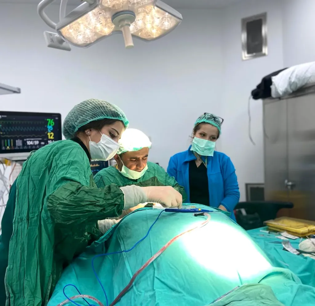
By Dr. Nejmi Öztürk, ophthalmologist and eye surgeon, at HG Hospital in Kahramanmaraş, Türkiye — dilated retinal examination, intravitreal injections, retinal laser and vitrectomy
- What the retina does and which diseases threaten vision
- Symptoms that need urgent attention: flashes of light, a sudden increase in floaters, a curtain coming down
- How the dilated eye examination, OCT and angiography are done
- Injections, laser and vitrectomy: which treatment for which disease
What is the retina, and what does it do?
The retina is the thin light-sensing layer that lines the back wall of the eye and converts light into nerve signals. It works like the film in a camera: light passing through the cornea and the lens is focused on the retina, the cells there convert the image into electrical signals, and the optic nerve carries these signals to the brain.
The small area at the center of the retina is called the macula; sharp-vision functions such as reading, recognizing faces and seeing colors depend on it. The peripheral parts of the retina are responsible for side vision and night vision. The fine blood vessels that supply the retina are directly affected by diabetes and high blood pressure; this is why a large share of retinal diseases are vascular diseases.
Retinal damage is usually irreversible; the aim of treatment is to stop the damage and preserve the remaining vision. Early diagnosis is therefore as important as the treatment itself.
Where is the retina, and what does it do?
Cross-section of the eye: the path of light and the position of the retina, the macula and the optic nerve.
- Cross-section of the eyeLight passes through the cornea and the lens, crosses the vitreous gel and falls on the retina, which lines the back wall of the eye. The retina converts this light into nerve signals, and the signals travel to the brain through the optic nerve.
What are the symptoms of retinal diseases?
Retinal diseases usually progress without pain, with a gradual and unnoticed deterioration of vision; people with diabetes and high blood pressure therefore need an examination without waiting for symptoms.
- Blurring in the center of vision, straight lines appearing bent or wavy (macular diseases)
- A dark or empty area in the middle of vision
- Reduced night vision, difficulty adapting to the dark (inherited diseases such as retinitis pigmentosa)
- Narrowing of side vision, seeing as if through a tunnel
- Colors appearing faded, reduced contrast
- Spots and threads floating in front of the eye (floaters)
Who should have regular retinal examinations?
People with diabetes and high blood pressure, everyone over 40, people with high myopia and those with a family history of retinal disease should have regular retinal examinations even if they have no symptoms.
| Who | Recommended frequency | Why |
|---|---|---|
| Type 2 diabetes | At diagnosis and then once a year | Diabetic retinopathy begins without symptoms |
| Type 1 diabetes | 5 years after diagnosis, then once a year | The risk increases with duration |
| Pregnant women with diabetes | At the start of pregnancy and every three months | Pregnancy can accelerate retinopathy |
| High blood pressure | Once a year | Vascular damage can be seen in the retina |
| Over 40 | Every 1–2 years | Macular degeneration and vascular occlusions increase with age |
| High myopia | Once a year | Risk of retinal thinning, tears and detachment |
| Family history of retinal disease | At the interval set by the physician | Early diagnosis of inherited diseases |
The pupil-dilating drops are not suitable for some patients: in people with a narrow-angle eye structure the drops can raise the eye pressure suddenly, so the anterior chamber angle is assessed before the drops are given. You cannot drive for a few hours after the drops; it is recommended not to come to the examination alone.
How is the retinal examination done, and which tests are used?
The retinal examination is done by looking at the back of the eye with a biomicroscope and special lenses after pupil-dilating drops have been applied; if considered necessary, detailed imaging with OCT and angiography is added.
- Dilated eye examination (with the pupil widened): The drops take effect in 20–30 minutes; the physician examines the retina, the blood vessels and the optic nerve directly. The effect lasts 4–6 hours; during this time near vision is blurred and light sensitivity is increased.
- OCT (optical coherence tomography): Takes cross-sectional images of the retinal layers at micron resolution. Macular edema (fluid build-up in the macula), macular holes and membrane formation are seen with this method. It does not touch the eye and takes a few minutes.
- Fluorescein angiography: A dye injected into a vein in the arm circulates through the retinal vessels while a series of photographs is taken; leaking and blocked vessels are seen. The dye can give the skin and urine a yellow tinge for a day; rarely an allergic reaction can occur.
- OCT angiography: Images the retinal vessels without dye; preferred for follow-up and in patients who cannot receive the dye.
- Eye ultrasound: When the retina cannot be seen because of a dense cataract or bleeding inside the eye, it shows whether the retina is in place.
- Visual field and Amsler tests: Loss of side vision and, in macular diseases, lines appearing bent are monitored with these tests.
How are retinal treatments applied?
There are three main treatments for retinal diseases: intravitreal injection, retinal laser and vitrectomy surgery. Which one is applied is determined by the disease, its stage and the imaging findings; in most patients these treatments are used together or one after another.
- Intravitreal injection (injection into the eye): After anesthetic drops, the surface of the eye is cleaned with an antiseptic; an anti-VEGF medicine or a steroid (cortisone) is delivered into the eye through the white of the eye with a very fine needle. The procedure takes a few minutes; patients describe a feeling of pressure. It is used for macular degeneration, diabetic macular edema and vascular occlusions; it is repeated at set intervals.
- Retinal laser (argon laser photocoagulation): Applied with anesthetic drops and a special lens, with the patient seated. The edges of retinal tears are sealed, and in diabetes the leaking or oxygen-starved areas are treated. It takes 10–20 minutes; there is brief stinging and a sensation of bright flashes.
- Vitrectomy: In the operating room, under local or general anesthesia, the gel inside the eye is removed through micro-incisions and the retina is repaired. It is performed for retinal detachment, advanced diabetic bleeding, macular holes and membrane formation; the details are explained on the vitrectomy page.
Which treatments are used in the retina?
Laser, intravitreal injection and surgery: three separate aims, shown schematically on a cross-section of the eye.
- Retinal laserThe laser burns the peripheral areas of the retina that lack oxygen in small spots, which reduces the signal for new vessel growth. The central vision area is spared.
- Intravitreal injectionAfter anesthetic drops, the medication is given into the vitreous cavity through the front side of the eye. It aims to reduce fluid build-up and the signal for new vessel growth.
- Surgery (vitrectomy)When there is bleeding, traction or a detachment, the eye is entered through three small ports, the vitreous is removed and the retina is repaired.
What happens after the examination and treatment?
Recovery after retinal treatments varies with the disease and the method; after an injection or laser you return to daily life the same day, whereas recovery after vitrectomy takes weeks.
- After the examination (day 1): The effect of the drops wears off towards the evening; do not drive; sunglasses bring relief.
- After an injection (week 1): There may be stinging and redness on the first day; the small blood spot at the injection site clears by itself. Increasing pain, redness and reduced vision are warning signs of infection; seek care immediately. Check-ups are usually every 4–8 weeks; whether to repeat the injection is decided with OCT.
- After laser (month 1): There may be slight blurring and light sensitivity for a few days; the effect settles within weeks and is assessed at the check-up.
- After vitrectomy: The positioning rules, the drop schedule and the recovery of vision over weeks to months are explained in detail on the vitrectomy page.
For patients coming from districts such as Elbistan, Afşin and Göksun, the examination, OCT and angiography are done on the same day as far as possible; repeat injections and check-ups are planned so that they are grouped into a single day.
What are the risks and limitations?
Retinal treatments aim to stop the damage; they cannot always restore lost vision, and each method has its own risks.
- Intravitreal injection: Rare but serious infection inside the eye, a temporary rise in eye pressure, bleeding, very rarely a retinal tear; the need to repeat the treatment.
- Retinal laser: Reduction in side vision and night vision (particularly with wide-area laser), temporary macular edema, small visual field losses near the laser scars.
- Vitrectomy: Cataract development, a rise in eye pressure, re-detachment, infection.
- General limitation: Diabetic retinopathy and macular degeneration are chronic diseases; treatment keeps progression under control, and management of blood sugar and blood pressure is part of the treatment.
Which treatment is used for which retinal disease?
The table below summarizes the common retinal diseases, their symptoms and the main treatment options; a link is given to the detailed page for each disease.
| Disease | Typical symptom | Main treatment options | Details |
|---|---|---|---|
| Diabetic retinopathy | No symptoms for a long time; at an advanced stage blurring, loss of vision due to sudden bleeding | Blood sugar control, anti-VEGF injections, retinal laser, vitrectomy at an advanced stage | Diabetic retinopathy |
| Macular degeneration (age-related macular degeneration) | Blurring in the center, lines appearing bent | In the dry type, follow-up and the supportive measures recommended by the physician; in the wet type, anti-VEGF injections | Macular degeneration |
| Retinitis pigmentosa (night blindness) | Night blindness, narrowing of side vision | Low-vision aids, genetic counseling, regular follow-up; treatments at the research stage are discussed without promises | Retinitis pigmentosa |
| Retinal tear and detachment | Flashes of light, an increase in floaters, a curtain coming down | Laser for a tear; for a detachment, surgical methods such as vitrectomy, gas and scleral buckling | Vitrectomy |
| Retinal vascular occlusion | Sudden reduction of vision in one eye | Anti-VEGF injections, laser; management of blood pressure and vascular risk factors | This page |
| Macular hole and epiretinal membrane | Distortion in the center, warped vision | Vitrectomy and membrane peeling | Vitrectomy |
Retinal examination and treatment in Kahramanmaraş
Retinal examination, assessment with OCT and angiography, intravitreal injections, retinal laser and vitrectomy are performed by Dr. Nejmi Öztürk, ophthalmologist and eye surgeon, at HG Hospital in Kahramanmaraş, Türkiye. Dr. Öztürk’s areas of interest include retinal diseases, vitrectomy and the diagnosis and treatment of retinopathy of prematurity (ROP) in premature babies.
HG Hospital is a private hospital contracted with the Turkish social security institution (SGK); patients with valid SGK coverage may use it, although a private-hospital co-payment may apply. Retinal treatments (anti-VEGF injections, laser, vitrectomy) are generally covered by SGK; for the injection medicines, SGK may require a medical board report. Patients without SGK coverage, such as international patients and visitors from abroad, pay privately or through their private or travel health insurance; the hospital checks coverage before treatment. Fee information is given by the hospital after the examination, and insurance coverage is clarified at the consultation.
Patients from Elbistan, Afşin, Göksun, Ekinözü and Nurhak come to HG Hospital in the center of Kahramanmaraş for examination and treatment; Dr. Öztürk does not see patients in Elbistan. With emergency symptoms, go to the nearest emergency department and ask for a same-day referral; for travel and planning, see the Elbistan and the surrounding area page.
Dr. Nejmi Öztürk sees patients at HG Hospital in Kahramanmaraş, Türkiye. For patients travelling from other districts or from abroad, examination, tests and treatment planning can be arranged for the same day.
Hospital switchboard (within Türkiye): 444 46 46 · E-mail: info@nejmiozturk.com · HG Hospital, Üngüt Mevkii, Prof. Dr. Necmettin Erbakan Blv. No:209, Onikişubat / Kahramanmaraş, Türkiye
Frequently asked questions
Why are drops used in the dilated eye examination, and how long does the effect last?
The drops widen the pupil so that the whole retina can be seen; without dilation only the center can be seen. The effect begins in 20–30 minutes and lasts 4–6 hours. During this time near vision is blurred and light is uncomfortable; you should not drive.
I have floaters; are they dangerous?
A few floating spots that have been present for a long time and do not change are usually due to age-related changes in the gel of the eye and are harmless. Floaters that begin suddenly and increase rapidly in number, especially if accompanied by flashes of light, can be a sign of a retinal tear. In that case a dilated eye examination is needed the same day.
What do flashes of light and the sensation of a curtain coming down mean?
Flashes of light occur when the gel of the eye pulls on the retina and can be a warning sign of a tear. A shadow or curtain coming down from one edge of the visual field can indicate that the retina has begun to separate. Both require an urgent examination; early treatment preserves vision.
I have diabetes; how often should I have a retinal examination?
The first examination is at diagnosis in type 2 diabetes and 5 years after diagnosis in type 1 diabetes; after that it is repeated once a year. If retinopathy is found, the physician shortens the interval to 3–6 months. During pregnancy the examinations are more frequent.
Is the eye injection painful, and how many times is it done?
Because the eye is numbed with drops, pain is not expected during the procedure; there is a feeling of pressure, and it lasts a few seconds. The number of injections depends on the disease: most patients have three monthly injections at the start, followed by repeat injections at intervals according to the OCT findings. Treatment is usually long-term.
Does retinal laser improve vision?
Retinal laser is done not to increase vision but to preserve the existing vision and to stop progression such as bleeding, leakage and detachment. Laser applied around a tear aims to prevent detachment. Vision usually stays the same; in some patients there may be some improvement as the edema decreases.
What is the difference between OCT and angiography?
OCT shows the retinal layers in cross-section; it detects structural problems such as edema, holes and membranes without dye. Angiography shows leakage from the vessels and blockages; in fluorescein angiography a dye is injected into a vein in the arm, whereas OCT angiography uses no dye. The two complement each other.
Are retinal diseases hereditary?
Some retinal diseases, such as retinitis pigmentosa, are hereditary and can be seen in more than one member of a family. Diabetic retinopathy and macular degeneration are not directly hereditary, although a family history increases the risk of macular degeneration. Regular examinations are recommended for people with a family history of retinal disease.
Are retinal treatments covered by SGK, and how much do they cost?
Anti-VEGF injections, retinal laser and vitrectomy are generally covered by SGK; the injection medicines may require a medical board report. Fee information is given by the hospital after the examination; insurance coverage is clarified at the consultation. A private-hospital co-payment may apply at HG Hospital.
I live in Elbistan; what should I do if I suddenly see flashes of light?
Sudden flashes of light, an increase in floaters or a curtain coming down is an emergency; see the nearest ophthalmologist or go to an emergency department the same day. Dr. Öztürk sees his patients only at HG Hospital in the center of Kahramanmaraş; the road from Elbistan to Kahramanmaraş takes about 2 hours. For planned examinations, the tests are completed on the same day as far as possible.
Sources
- Turkish Ophthalmological Society — todnet.org
- American Academy of Ophthalmology (AAO) — aao.org
- National Eye Institute (NEI) — nei.nih.gov
Related pages
This page is for information only; diagnosis and treatment decisions are made after an examination. The results of any surgical or interventional procedure may vary from person to person. You are advised to obtain detailed advice from your physician before any procedure.

Ophthalmologist and eye surgeon. Graduate of Erciyes University Faculty of Medicine; completed his residency at Ankara Atatürk Training and Research Hospital. With 21 years of experience in cataract and refractive surgery, strabismus, glaucoma, retina and oculoplastic surgery, he sees patients at HG Hospital in Kahramanmaraş, Türkiye. Biography and training
Dr. Nejmi Öztürk
Ophthalmologist and eye surgeon
HG Hospital, Üngüt Mevkii, Prof. Dr. Necmettin Erbakan Blv. No:209, Onikişubat / Kahramanmaraş
+90 506 377 47 68WhatsAppAppointment formHospital switchboard (within Türkiye): 444 46 46 · info@nejmiozturk.com
- Private patients and private health insurance are welcome; patients with SGK coverage may use it (conditions are confirmed by the hospital)
- Same-day examination and test planning for patients travelling from other districts or from abroad
- Getting here and patient information
- Patients from Elbistan and the region