
Uveitis and Diseases of the Uvea (Inflammation Inside the Eye)
Diagnosis, treatment and regular follow-up of anterior, intermediate, posterior and panuveitis by Dr. Nejmi Öztürk, ophthalmologist and eye surgeon, at HG Hospital in Kahramanmaraş, Türkiye
- What the uvea is; the difference between anterior, intermediate and posterior uveitis and panuveitis
- Symptoms: pain, light sensitivity, redness, blurred vision, floaters
- The link with Behçet’s disease and rheumatic conditions; diagnostic tests and working together with rheumatology
- Cortisone drops, injections, systemic and biological treatments; complications and follow-up
What is the uvea, and what is uveitis?
The uvea is the middle layer of the eye, rich in blood vessels, and it has three parts: at the front the iris (the colored part of the eye), in the middle the ciliary body (the structure that produces the fluid inside the eye and focuses the lens), and at the back the choroid (the vascular layer that nourishes the retina). Uveitis is inflammation of this layer; it is often called “eye inflammation”, but unlike conjunctivitis (pink eye) it affects the inside of the eye and can lead to permanent loss of vision if it is not treated.
Uveitis is divided into four groups according to where the inflammation is; the location determines both the symptoms and how intensive the treatment needs to be:
| Type | Part affected | Typical features |
|---|---|---|
| Anterior uveitis (iritis, iridocyclitis) | The iris and the front part of the ciliary body | The most common type; it starts with sudden pain, redness and light sensitivity and usually responds well to drop treatment. |
| Intermediate uveitis | The vitreous (the gel inside the eye) and the back part of the ciliary body | There is little pain; floaters and blurring are the main complaints. It can occur in young people and together with multiple sclerosis. |
| Posterior uveitis | The choroid and the retina | Usually causes no pain, but it is the type that threatens vision the most; infections such as toxoplasmosis and Behçet’s disease are the main causes. |
| Panuveitis | The whole uvea | Anterior and posterior findings occur together; seen in Behçet’s disease, sarcoidosis and Vogt-Koyanagi-Harada disease. |
What are the symptoms of uveitis, and who gets it?
The most common symptoms of uveitis are eye pain, not being able to tolerate light, redness and blurred vision. The symptoms vary according to the part affected:
- A deep, throbbing pain in the eye and an ache radiating above the brow
- Light sensitivity (photophobia); squinting in bright light, increased tearing
- Redness: Unlike conjunctivitis, it is most pronounced in a ring around the colored part of the eye, and there is no discharge
- Blurred or hazy vision
- Floaters (spots drifting across the vision); the main complaint in intermediate and posterior uveitis
- A small or irregularly shaped pupil (because the iris has stuck down)
- Recurrent red eye: Redness and pain recurring in the same eye every few months suggest uveitis
What causes uveitis?
In a large proportion of uveitis cases, the cause is the immune system targeting the eye; in some there is an infection, and in the rest no cause is found despite all investigations (idiopathic uveitis).
- Autoimmune and rheumatic diseases: Behçet’s disease (common in Türkiye; it causes posterior uveitis and inflammation of the retinal blood vessels), ankylosing spondylitis and HLA-B27-related diseases (recurrent anterior uveitis), juvenile idiopathic arthritis in children (a silent, insidious anterior uveitis), rheumatoid arthritis, sarcoidosis, inflammatory bowel disease, multiple sclerosis.
- Infections: Toxoplasmosis (transmitted through cat feces and undercooked meat), herpes and shingles viruses, tuberculosis, syphilis.
- Other: Eye injury, previous eye surgery.
Who is standard cortisone treatment not suitable for?
Uveitis treatment is not the same for everyone; in some situations cortisone on its own is not suitable and may even be harmful:
- Uveitis caused by infection: In uveitis due to toxoplasmosis, herpes or tuberculosis, cortisone is given only together with the medicine for the infection; used on its own, it makes the infection flare up.
- People whose eye pressure rises in response to cortisone: Eye pressure is monitored closely during treatment; if necessary, a switch is made to non-cortisone medicines.
- Uncontrolled diabetes, severe osteoporosis, stomach ulcer, active infection: The dose of oral or intravenous cortisone is limited; immunosuppressive medicines are brought forward.
- Pregnancy and breastfeeding: Some immunosuppressive medicines cannot be used; treatment is planned together with the obstetrician.
How are the examination and tests done?
Uveitis is diagnosed when inflammatory cells are seen inside the eye at the slit-lamp (biomicroscope) examination; finding the cause requires blood tests and, when needed, a rheumatology opinion. Dr. Nejmi Öztürk does the following at the examination:
- Slit-lamp examination: The cells and haze in the anterior chamber are graded; deposits behind the cornea and adhesions of the iris (synechiae) are looked for.
- Eye pressure measurement: Uveitis can both lower and raise the eye pressure; it is measured at every check-up.
- Retinal (fundus) examination: The pupil is dilated with drops and the vitreous, retina and choroid are examined.
- OCT (optical coherence tomography): Measures fluid build-up in the center of vision (macular edema); used to monitor the response to treatment.
- Fundus fluorescein angiography (eye angiogram): Shows inflammation and leakage in the retinal blood vessels; needed especially in Behçet’s disease.
- Blood and imaging tests: Full blood count, sedimentation rate, HLA-B27, syphilis and tuberculosis tests, a chest X-ray; toxoplasmosis and viral tests depending on the suspicion.
- Working with rheumatology: You are asked about joint, mouth ulcer, skin and bowel symptoms; when a systemic disease is suspected, the assessment is made together with rheumatology.
What happens in uveitis?
Inflammation of the vascular middle layer of the eye, and the signs that can be seen from the outside.
- The inflamed layerThe uvea is the vessel-rich middle layer of the eye. When it becomes inflamed there is pain, sensitivity to light, redness and blurred vision; the signs can be in the front, middle or back part of the eye.
- The aim of treatmentTreatment aims to suppress the inflammation and prevent lasting damage. It starts with drops; if needed, systemic treatment is added and an underlying disease is looked for.
How is uveitis treated?
The aim of uveitis treatment is to bring the inflammation under control quickly, to protect vision and to prevent recurrences. Treatment proceeds step by step; the location, severity and cause of the inflammation determine which step to start from:
- Cortisone (steroid) eye drops: The main treatment in anterior uveitis. At the start they are instilled frequently (sometimes every hour) and, as the inflammation settles, they are reduced step by step over weeks. Stopping them suddenly makes the inflammation flare up.
- Pupil-dilating drops (cycloplegics): They relieve pain by releasing the spasm of the ciliary muscle and prevent the iris from sticking to the lens; they temporarily blur near vision.
- Cortisone injection around or inside the eye: Used in intermediate and posterior uveitis and in macular edema. It is done in a few minutes under drop anesthesia; you feel pressure during the injection. The effect lasts from weeks to months.
- Oral or intravenous cortisone: A short course at a high dose is used in severe or posterior uveitis and when both eyes are affected; because of its side effects it is planned as a bridging treatment.
- Immunosuppressive medicines: In cases where cortisone cannot be stopped or which recur often, medicines such as methotrexate, azathioprine, mycophenolate and cyclosporine are started together with rheumatology; regular blood tests are needed.
- Biological treatments: In Behçet’s disease and resistant uveitis, anti-TNF biological agents or interferon are used by joint decision with rheumatology.
- Treatment of infection: If the cause is toxoplasmosis, herpes or tuberculosis, the appropriate antibiotic or antiviral medicine is given together with cortisone.
- Treatment of complications: Cataract or glaucoma that develops is treated surgically once the inflammation has been quiet for at least 3 months; permanent clouding of the vitreous may require a vitrectomy.
What are recovery and follow-up like?
In anterior uveitis, pain and light sensitivity usually decrease within a few days of starting drop treatment; it takes 4–6 weeks for the inflammation to settle completely. In posterior uveitis and in cases linked to a systemic disease, treatment lasts months, sometimes years.
- Week 1: The drops are used at frequent intervals; a check-up is done within 3–7 days and the eye pressure is measured. Sunglasses reduce light sensitivity.
- Month 1: As the cells in the anterior chamber decrease, the dose of drops is lowered step by step. The tapering schedule is given in writing; do not stop the drops on your own.
- Months 3–6 and beyond: Check-ups continue even after the inflammation has settled; in people on systemic medicines, blood tests and rheumatology check-ups are repeated. In silent childhood uveitis, regular examination is essential even without symptoms.
For patients coming from districts such as Elbistan, Afşin and Göksun, the intervals between check-ups are extended once the inflammation has settled, taking the travel into account; blood tests can be done at a center near home and the results assessed at the check-up.
What are the risks, complications and limitations?
The main risk of uveitis is that it leaves permanent damage in the eye when it is not treated or when it recurs. The treatment itself also has side effects, and follow-up is done to manage these two risks together.
- Cataract (clouding of the lens): Both the inflammation itself and long-term cortisone use cloud the lens; surgery is planned while the inflammation is quiet (cataract surgery).
- Glaucoma (high eye pressure): Inflammatory cells can block the outflow of fluid from the eye; cortisone also raises the pressure in some people (glaucoma treatment).
- Macular edema: Fluid build-up in the center of vision is the most common cause of vision loss in uveitis; it is monitored with OCT and may need an injection.
- Adhesions of the iris (synechiae): They distort the shape of the pupil and can lead to an eye pressure crisis.
- Clouding of the vitreous, retinal detachment, calcium deposits on the cornea (band keratopathy)
- Side effects of treatment: With systemic cortisone, weight gain, raised blood sugar and bone loss; with immunosuppressive medicines, susceptibility to infection and disturbances of liver function and blood counts. Regular blood tests are done for this reason.
- Recurrence: Uveitis linked to HLA-B27 and Behçet’s disease in particular runs in attacks; treatment aims to reduce the attacks and cannot promise to eliminate them completely.
Comparison of treatment options
Rather than being alternatives to one another, the treatment options are steps that are added to each other according to the severity of the disease.
| Option | When | Pros | Cons |
|---|---|---|---|
| Cortisone drops | Anterior uveitis | Effect limited to the eye, easy to use | Do not reach the back of the eye; eye pressure, cataract |
| Injection around / inside the eye | Intermediate and posterior uveitis, macular edema | Strong local effect, few side effects on the body | May need repeating; eye pressure, rarely infection |
| Systemic cortisone | Severe uveitis affecting both eyes | Fast and strong effect | Serious side effects on the body with long-term use |
| Immunosuppressive medicines | Chronic, recurrent uveitis | Reduce the need for cortisone | Effect starts after weeks; blood tests needed |
| Biological treatments | Behçet’s disease, resistant uveitis | Effective in resistant cases | Infection screening and rheumatology follow-up are mandatory |
Retinal problems caused by uveitis are described on the retinal diseases page, and the operation performed for permanent clouding of the vitreous on the vitrectomy page.
Diagnosis and follow-up of uveitis in Kahramanmaraş
The diagnosis and follow-up of uveitis and diseases of the uvea are carried out by Dr. Nejmi Öztürk at HG Hospital in Kahramanmaraş, Türkiye; the slit-lamp examination, retinal examination, OCT and the necessary blood tests are planned at the same hospital. When a systemic disease is suspected, a joint assessment is made with rheumatology and internal medicine.
Uveitis examinations, drop and injection treatments and surgery for complications are generally covered by SGK. HG Hospital is a private hospital contracted with the Turkish social security institution (SGK); patients with valid SGK coverage may use it, although a private-hospital co-payment may apply. Biological medicines may require a medical board report in Türkiye. Patients without SGK coverage, such as international patients and visitors from abroad, pay privately or through their private or travel health insurance; the hospital checks coverage before treatment. Fee information is given by the hospital after the examination, and insurance coverage is clarified at the consultation.
Patients from Elbistan, Afşin, Göksun, Ekinözü and Nurhak are examined at HG Hospital in Kahramanmaraş; Dr. Öztürk does not see patients in Elbistan. Do not put off the examination if you have a sudden painful red eye; for travel planning, see the patients from Elbistan and the surrounding area page.
Dr. Nejmi Öztürk sees patients at HG Hospital in Kahramanmaraş, Türkiye. For patients travelling from other districts or from abroad, examination, tests and treatment planning can be arranged for the same day.
Hospital switchboard (within Türkiye): 444 46 46 · E-mail: info@nejmiozturk.com · HG Hospital, Üngüt Mevkii, Prof. Dr. Necmettin Erbakan Blv. No:209, Onikişubat / Kahramanmaraş, Türkiye
Frequently asked questions
Is uveitis contagious?
No. Uveitis is inflammation of the inner layer of the eye and does not pass from person to person. It should not be confused with conjunctivitis (pink eye), which is contagious; in uveitis there is no discharge, and pain and light sensitivity are the main features.
Does uveitis go away completely?
An attack of anterior uveitis usually heals completely within 4–6 weeks with treatment. If there is an underlying disease such as Behçet’s disease or HLA-B27, attacks may recur; in that case treatment aims to make the attacks less frequent and to protect vision. Regular follow-up allows recurrences to be caught early.
When should I stop the cortisone drops?
The drops are reduced step by step over weeks and stopped after the inflammatory cells have disappeared at the examination. Stopping them suddenly on your own makes the inflammation flare up. Your doctor gives you the tapering schedule in writing.
How is eye involvement recognized in Behçet’s disease?
In Behçet’s disease, eye involvement usually begins with blurring of vision without pain, floaters or recurrent redness. Everyone with mouth and genital ulcers is advised to have an eye examination even without symptoms; inflammation of the retinal blood vessels can progress silently.
Is uveitis treatment covered by SGK?
Uveitis examinations, drop and injection treatments and operations for complications are generally covered by SGK; a private-hospital co-payment may apply. Biological medicines may require a medical board report in Türkiye. Patients without SGK coverage pay privately or through their private or travel health insurance; coverage conditions are clarified at the examination.
How much does uveitis treatment cost?
Fee information is given by the hospital after the examination; SGK and private insurance coverage is clarified at the consultation. Because the treatment step (drops, injection, systemic medicine) is decided at the examination, it is not possible to give information in advance.
Can children get uveitis?
Yes. In children with juvenile idiopathic arthritis, uveitis runs silently without pain or redness; for this reason children being followed by rheumatology should be brought for regular eye examinations even when they have no symptoms. Dr. Öztürk also works in pediatric ophthalmology.
Where should I go for a uveitis examination in Kahramanmaraş?
Dr. Nejmi Öztürk carries out the diagnosis and follow-up of uveitis at HG Hospital in Onikişubat. Patients with a sudden painful red eye and light sensitivity should not delay the examination. Appointment details are on the contact page.
Sources
- Turkish Ophthalmological Society — todnet.org
- American Academy of Ophthalmology (AAO) — aao.org
- National Eye Institute (NEI) — nei.nih.gov
Related pages
This page is for information only; diagnosis and treatment decisions are made after an examination. The results of any surgical or interventional procedure may vary from person to person. You are advised to obtain detailed advice from your physician before any procedure.
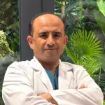
Ophthalmologist and eye surgeon. Graduate of Erciyes University Faculty of Medicine; completed his residency at Ankara Atatürk Training and Research Hospital. With 21 years of experience in cataract and refractive surgery, strabismus, glaucoma, retina and oculoplastic surgery, he sees patients at HG Hospital in Kahramanmaraş, Türkiye. Biography and training
Dr. Nejmi Öztürk
Ophthalmologist and eye surgeon
HG Hospital, Üngüt Mevkii, Prof. Dr. Necmettin Erbakan Blv. No:209, Onikişubat / Kahramanmaraş
+90 506 377 47 68WhatsAppAppointment formHospital switchboard (within Türkiye): 444 46 46 · info@nejmiozturk.com
- Private patients and private health insurance are welcome; patients with SGK coverage may use it (conditions are confirmed by the hospital)
- Same-day examination and test planning for patients travelling from other districts or from abroad
- Getting here and patient information
- Patients from Elbistan and the region