
Q-LASIK Eye Surgery
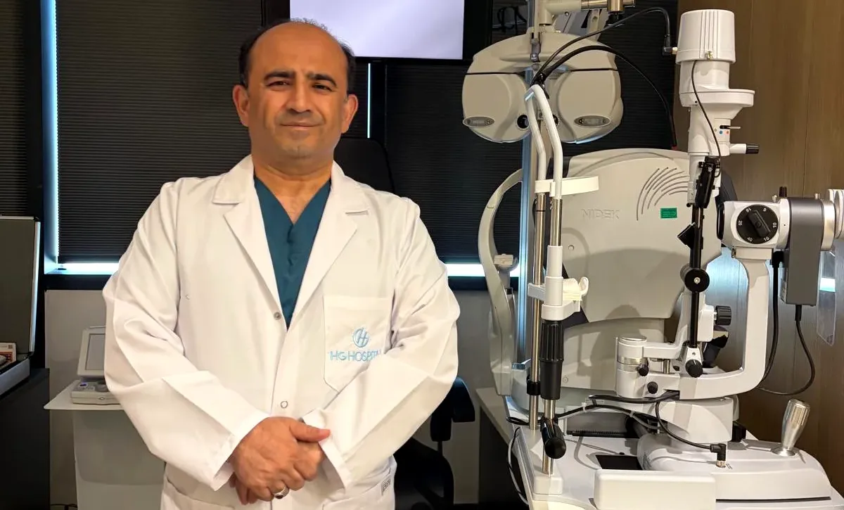
With Dr. Nejmi Öztürk, ophthalmologist and eye surgeon, at HG Hospital in Kahramanmaraş, Türkiye — Q-LASIK suitability assessment, flap creation with the Med Logics ML7 LASIK system and planning supported by RLES AI
- What Q-LASIK is — where the name comes from and which step sets the method apart
- The Med Logics ML7 LASIK system — timing, suction level, deviation in flap thickness, hinge orientation
- Why Q-LASIK comes up in thin corneas
- Examination and measurements: why the front surface alone is not enough
- RLES AI as a second check on the plan
- Recovery, risks and how the methods compare
What is Q-LASIK?
Q-LASIK is a LASIK procedure whose name comes from “Quick Visual Recovery”. Like every method in the LASIK family it has two stages: a thin flap is prepared on the front of the cornea (the clear front layer of the eye), the flap is lifted, and the stromal tissue beneath it is reshaped with the excimer laser by the amount of the prescription. The flap is then laid back in place and no stitches are needed.
What distinguishes the method is not the excimer stage but the way the flap is created. In the LASIK family it is the flap stage that shapes the outcome most directly: how long it takes, at what suction level, at what thickness and in which direction the flap is prepared determines both the comfort of the procedure and the speed of recovery. In Q-LASIK this stage is carried out with the Med Logics ML7 LASIK system.
The ablation stage — shaping the tissue with the laser — is then planned with the asphericity of the cornea (its Q value) taken into account. The aim is to use tissue efficiently while correcting myopia (nearsightedness), hyperopia (farsightedness) and astigmatism in eyes that are suitable.
The Med Logics ML7 LASIK system and the flap stage
Med Logics ML7 is an FDA- and CE-approved LASIK system that prepares the flap under autonomous control. Its main technical features at the flap stage are as follows:
- About 7 seconds, low suction: The flap is created in roughly 7 seconds including the suction phase, at a low suction level of 550 mmHg. The eye stays under suction for no longer than this period, which adds to the comfort of the procedure and reduces corneal swelling.
- Close to the intended thickness (SD ±5 µm): The deviation between the planned and the achieved flap thickness is very small. That makes it possible to calculate the amount of tissue that will remain in the cornea after the laser with greater confidence before surgery.
- A planar flap, cuttable along any axis: The flap can be cut temporally (towards the temple), nasally, superiorly, or at any degree chosen according to the astigmatic axis. The cut is planar, so the thickness is close to uniform across the whole flap.
- What a temporal hinge means: The sensory nerves of the cornea enter mainly at the 3 and 9 o’clock positions. A temporal flap preserves part of these fibres, so the corneal reflex returns sooner and complaints of dryness after surgery tend to be fewer.
- An eyelid speculum is not obligatory: Because a speculum is not required at the flap stage, the procedure is easier to carry out in patients with a narrow lid opening, and the risk of losing suction is reduced.
- Less corneal swelling: Working briefly and at low pressure limits the swelling that forms in the cornea, which is why visual recovery completes within the same day.
This is where the difference the patient notices comes from: burning and stinging after surgery are markedly less pronounced, and vision sharpens within a short time.
What is the difference between Q-LASIK and standard LASIK?
| Comparison | Q-LASIK (Med Logics ML7) | Standard LASIK |
|---|---|---|
| Flap time | About 7 seconds including suction | Longer, depending on the system |
| Suction level | 550 mmHg (low) | Usually higher |
| Thickness deviation | SD ±5 µm | Varies by system |
| Hinge orientation | Selectable along any axis; temporal can be chosen | Whichever direction the system allows |
| Corneal swelling | Limited | Can be greater |
| Visual recovery | Sharpens within 5–6 hours | Usually the following day |
These differences do not mean the method will give a better outcome in everyone. Which method suits you is determined by assessing the full set of measurements.
Who is it assessed for?
- People aged 18 or over whose prescription has not changed appreciably over the past 12 months
- Those whose myopia, hyperopia and astigmatism fall within a suitable range
- Those with a regular corneal topography and an adequate tear film
- Eyes whose corneal thickness is borderline for standard LASIK but whose topography is regular
- People for whom returning to ordinary life the same day matters
Who is it not suitable for?
- Anyone with signs of keratoconus or corneal thinning (ectasia) — no corneal laser, Q-LASIK included, is applied to these eyes
- Those whose corneal thickness falls below the limit
- Those with an active eye infection, untreated dry eye or a history of ocular herpes (cold sore virus)
- Those with uncontrolled diabetes and certain rheumatic diseases
- Women who are pregnant or breastfeeding
- Those in whom a cataract has developed — laser is of no benefit here, and lens surgery is assessed instead
Age-related difficulty with near vision (presbyopia) calls for separate planning; presbyopia protocols are assessed in suitable cases.
Thin corneas and Q-LASIK
Because the flap can be planned thin and the deviation in its thickness is small, more tissue is left behind in the cornea. For that reason Q-LASIK can be considered as an option in some eyes where standard LASIK cannot be applied on account of a thin cornea.
Thickness on its own, however, is not a sufficient criterion. The decision is made by weighing corneal topography, tomography, the thickness map and the calculation of the tissue that will remain after the laser together. In eyes found unsuitable, surface methods that require no flap, or intraocular lens options, come into consideration.
Examination and measurements: why is the front surface alone not enough?
Because a flap is lifted from the cornea in Q-LASIK, the measurements taken before surgery are as decisive as the method itself. Looking at the front surface of the cornea alone is not enough: when a cornea begins to weaken, the change appears on the back surface before anything else, while the front surface can still look normal for some time. That is why a tomographic assessment is carried out, showing the front surface, the back surface and the thickness at every point together.
- Prescription measurement with and without drops
- Corneal topography and tomography (front surface, back surface, thickness map)
- Assessment of eye pressure and the anterior chamber angle
- Tear tests and examination of the ocular surface
- Measurement of pupil diameter in the dark
- Retinal examination with the pupil widened by drops
Contact lens wear can temporarily distort the shape of the cornea and mislead the measurements. Soft lens wearers are therefore asked to leave their lenses out for at least a week before the examination, and rigid lens wearers for longer.
Checking the plan with RLES AI
In laser planning, Dr. Nejmi Öztürk uses RLES AI (Refractive Laser Eye Surgery Artificial Intelligence) as a second layer of checking — explainable artificial-intelligence-based clinical decision support software built for refractive surgery. The software does not diagnose, does not decide whether to operate and does not guarantee an outcome; what it does is recalculate the measurements taken at the examination independently, so that what can be known is not overlooked. Because it is explainable, every warning it raises shows which measurement and which calculation it rests on — so the surgeon can verify each one, and you can see what the plan was based on.
- Tissue safety calculations: The thickness that will remain in the cornea after treatment is calculated and compared against safety floors.
- Device nomogram check: The manufacturer nomograms of the technologies used in laser eye surgery are verified against the patient’s own measurements.
- Ectasia and keratoconus screening: Tomographic indices are read together, and indices that depend on one another are not counted as separate evidence, so both false alarms and false reassurance are reduced.
- Contact-lens-related surface change: Temporary distortion that could mislead the measurements is flagged.
- Dry eye, eye pressure and the anterior chamber angle are checked separately in every plan.
- A missing measurement is not hidden: If an item has not been measured, it is reported by name; “unknown” is never treated as “normal”.
A single laser plan involves close to three hundred items that have to be queried, read and calculated across both eyes. Most of them are items the surgeon already knows; the hard part is not knowing them but querying every one of them, in the same order and without exception, in every case. RLES AI puts that check on record. The final clinical decision rests with the surgeon and is not made without an examination.
How is the surgery performed?
- Anesthetic drops: Numbing drops are instilled into the eye. No needle is used and no general anesthesia is needed. You do not feel pain during the procedure; a sense of touch and pressure is normal.
- The flap with Med Logics ML7: A thin flap is created on the front of the cornea in about 7 seconds including suction, and is lifted from one edge. There is a sensation of pressure for a few seconds at this stage, and vision dims briefly.
- Laser application: The excimer laser shapes the cornea by removing the planned amount of tissue. You are asked to look at a light while this happens; an eye-tracking system keeps the laser aligned through small movements.
- Closing: The flap is settled back into its own bed; no stitches are placed and it adheres by itself. For both eyes the procedure takes about 10–15 minutes including preparation.
Q-LASIK step by step
The flap prepared with the Med Logics ML7 LASIK system and the excimer laser stages, shown schematically on a cross-section of the cornea.
- Preparing the flapAfter anaesthetic drops, the Med Logics ML7 LASIK system prepares a thin flap in the front part of the cornea with low suction, in about 7 seconds.
- Lifting the flapThe flap is lifted gently on its hinge, opening the stromal tissue underneath for treatment. The flap is never detached completely.
- Correcting the refractive errorGuided by the plan derived from the measurements, the excimer laser removes tissue from the stromal surface at micron level and changes the curvature of the cornea.
- Replacing the flapThe flap is laid back and adheres on its own; no stitches are used. The whole procedure takes about 10–15 minutes for both eyes.
EpitheliumStromaArea the laser acts on
The recovery process
| Period | What to expect | What to do |
|---|---|---|
| Immediately after surgery | Vision approaches 80–90% of the targeted level; mild blurring is normal | Protective glasses are worn; travel home with a companion and do not drive |
| Within 5–6 hours | Watering and light sensitivity may occur; by the end of this period vision sharpens | Rest or sleep; do not rub the eye |
| The next day | Most patients return to their daily work and to driving | Follow-up examination; use the drops regularly |
| Week 1 | A mild sensation of dryness may be present | Keep water out of the eye and avoid rubbing; showering from day 3 onwards |
| Weeks 1–4 | Night glare and dryness gradually subside | No make-up for 1–2 weeks; no swimming pools or sweaty sport for 3–4 weeks |
| Month 1 and beyond | Visual acuity settles | Month 1 follow-up |
For patients travelling in from out of town, the procedure can be scheduled for the morning; once the checks are complete, travelling the same day is unproblematic for most patients. That decision is made according to the examination findings.
Risks and limitations
- Temporary dry eye: Less likely with Q-LASIK thanks to the temporal hinge, but it can still occur; it is managed with artificial tears.
- Night glare and halos: These can persist through the early months, usually up to around the third month.
- Light sensitivity: Daylight can seem too bright in the early period.
- Flap-related problems: Epithelial ingrowth, or displacement of the flap through unintentional contact during the opening day or two, is uncommon; it can be corrected with early intervention.
- Infection: Can occur if the drops are not used regularly and hygiene rules are not followed.
- An outcome different from the target: Particularly with high prescriptions, a small amount of prescription can remain and additional correction may be needed.
- Ectasia related to corneal biomechanics: A rare but serious condition that has been reported at a later stage; in borderline eyes the method is changed or surgery is not recommended.
- Presbyopia: Q-LASIK corrects distance vision; it does not remove the age-related change in near vision.
The laser corrects the prescription as it stands on the day of surgery. In a healthy eye the outcome is lasting; the eye nonetheless ages along with the body, and systemic conditions such as diabetes and hypertension can affect the prescription again later on. No refractive treatment can carry a lifetime guarantee.
Q-LASIK, No-Touch or SMILE?
- Q-LASIK: The flap is prepared with Med Logics ML7. Vision sharpens the same day and discomfort in the early days is limited. It can be applied in myopia, hyperopia and astigmatism.
- No-Touch (Trans-PRK): There is no flap and no instrument touches the corneal surface. It is preferred in patients whose eyes are not suited to the LASIK family; the early days are more uncomfortable and sharpening takes weeks.
- SMILE: There is no flap; a disc of tissue is removed through a 2–4 mm incision. It is applied in myopia and myopic astigmatism.
In patients who have had retinal detachment surgery, who have raised eye pressure or who present some other particular circumstance, surface methods may have to be preferred. A detailed comparison of the methods is on the laser eye surgery page.
Q-LASIK assessment in Kahramanmaraş
Q-LASIK suitability assessment, the measurements and the surgery are carried out at Private HG Hospital in Kahramanmaraş, Türkiye, by Dr. Nejmi Öztürk, ophthalmologist and eye surgeon. For patients travelling in from Elbistan, Afşin, Göksun, Ekinözü and Nurhak, the examination and tests are grouped into the same day as far as possible; the surgery is scheduled for a separate day, since it requires a break from contact lenses and some preparation. To make an appointment, you can use the details on the contact page.
In eyes where laser is found unsuitable, ICL (intraocular lens) and lens surgery are assessed; if a progressive irregularity is detected on the corneal map, keratoconus treatment is planned beforehand.
Dr. Nejmi Öztürk sees patients at HG Hospital in Kahramanmaraş, Türkiye. For patients travelling from other districts or from abroad, examination, tests and treatment planning can be arranged for the same day.
Hospital switchboard (within Türkiye): 444 46 46 · E-mail: info@nejmiozturk.com · HG Hospital, Üngüt Mevkii, Prof. Dr. Necmettin Erbakan Blv. No:209, Onikişubat / Kahramanmaraş, Türkiye
Frequently asked questions
What does the “Q” in Q-LASIK stand for?
The abbreviation Q-LASIK stands for “Quick Visual Recovery”. Immediately after surgery, vision approaches 80–90% of the targeted level, and it sharpens within 5–6 hours.
What is the difference between Q-LASIK and standard LASIK?
The essential difference is the way the flap is created. With the Med Logics ML7 LASIK system the flap is prepared in about 7 seconds including suction, under low suction of 550 mmHg, and very close to the intended thickness (SD ±5 µm). The hinge orientation can also be selected along any axis. The ablation, meanwhile, is planned with corneal asphericity taken into account.
How long does the flap stage take?
About 7 seconds, including the time suction is applied. The eye stays under suction for no longer than that period. For both eyes together, the procedure takes about 10–15 minutes including preparation.
Is pain felt during the surgery?
Because anesthetic drops are used, no pain is felt during the procedure; a sense of touch and pressure is normal. Watering and stinging may occur in the early hours and pass quickly.
When can I go back to my normal life?
Vision sharpens within 5–6 hours, and most patients return to their normal life at home the same day. Do not drive on the day of surgery; the next day you can go to work and drive. Showering is allowed from day 3, make-up after 1–2 weeks, and swimming pools and sweaty sport after 3–4 weeks.
My cornea is thin — can I have Q-LASIK?
Because a thin flap with little deviation leaves more tissue in the cornea, Q-LASIK can be assessed in some eyes where standard LASIK cannot be applied. Thickness alone is not the criterion, though; topography, tomography and the calculation of remaining tissue are weighed together.
Can I have Q-LASIK if I have keratoconus?
No. In eyes with signs of keratoconus or corneal thinning, no corneal laser is applied, Q-LASIK included. In these eyes keratoconus treatment comes beforehand, and in suitable cases the intraocular lens (phakic ICL) option is assessed.
Why is more than one corneal map taken at the examination?
When a cornea begins to weaken, the change usually appears on the back surface first; the front surface can still look normal for some time. The front surface, the back surface and the thickness map are therefore read together — one can look normal while another already shows an early sign.
Does artificial intelligence make the decision to operate?
No. RLES AI does not diagnose, does not decide whether to operate and does not guarantee an outcome. It checks the tissue safety calculations, the nomogram values of the devices in use and the indicators of ectasia risk, and reports any missing measurement by name. Your doctor makes the decision.
Is the outcome permanent for life?
The laser corrects the prescription as it stands on the day of surgery, and in a healthy eye the outcome is lasting. The eye ages too, however, and systemic conditions can affect the prescription later on. No refractive treatment can carry a lifetime guarantee.
Sources
- Lopes BT, Ramos IC, Salomão MQ, et al. Enhanced Tomographic Assessment to Detect Corneal Ectasia Based on Artificial Intelligence. American Journal of Ophthalmology. 2018;195:223-232.
- Turkish Ophthalmological Society — todnet.org
- American Academy of Ophthalmology (AAO) — aao.org
- National Eye Institute (NEI) — nei.nih.gov
Related pages
- Laser eye surgery — general information
- No-Touch (Trans-PRK)
- PRK
- Wavefront- and topography-guided laser
- ICL (intraocular lens)
This page is for informational purposes; diagnosis and treatment decisions are made after an examination. The results of any surgical or interventional procedure may vary from person to person. You are advised to obtain detailed advice from your physician before any procedure.
Ophthalmologist and eye surgeon. Graduate of Erciyes University Faculty of Medicine; completed his residency at Ankara Atatürk Training and Research Hospital. With experience in ophthalmology since 2005 in cataract and refractive surgery, strabismus, glaucoma, retina and oculoplastic surgery, he sees patients at HG Hospital in Kahramanmaraş, Türkiye. Biography and training
Dr. Nejmi Öztürk
Ophthalmologist and eye surgeon
HG Hospital, Üngüt Mevkii, Prof. Dr. Necmettin Erbakan Blv. No:209, Onikişubat / Kahramanmaraş
+90 506 377 47 68WhatsAppAppointment formHospital switchboard (within Türkiye): 444 46 46 · info@nejmiozturk.com
- Private patients and private health insurance are welcome; patients with SGK coverage may use it (conditions are confirmed by the hospital)
- Same-day examination and test planning for patients travelling from other districts or from abroad
- Getting here and patient information
- Patients from Elbistan and the region