
Keratoconus Treatment
With Dr. Nejmi Öztürk, ophthalmologist and eye surgeon, at HG Hospital in Kahramanmaraş, Türkiye — diagnosis, corneal follow-up and planning of the treatment step that suits you
- What keratoconus is, at what age it begins and why it progresses
- Why a constantly increasing glasses prescription and eye rubbing are warning signs
- Which measurements are used for diagnosis and how the stages are determined
- The treatment steps from glasses to corneal transplant, and the follow-up plan
What is keratoconus?
Keratoconus is the progressive thinning of the cornea, the clear front layer of the eye, and its bulging forward into a cone shape. A healthy cornea is a smooth dome; it bends light evenly and focuses it sharply on the retina. In keratoconus this dome weakens in one area and bulges outward, so light is bent irregularly. The result is blurred and distorted vision that cannot be fully corrected with glasses.
The disease usually begins in adolescence and can progress until the 30s. Progression is faster in young people; after the age of 35–40 it usually slows down, because the cornea stiffens naturally. Keratoconus usually affects both eyes, but the two eyes can be at different stages.
Keratoconus is not an inflammatory disease and is not contagious. As corneal imaging methods have become widespread, the number of patients diagnosed at an early stage has increased. In a press statement in June 2026, Dr. Nejmi Öztürk informed the public about the symptoms of keratoconus and the importance of early diagnosis.
What are the symptoms of keratoconus?
The most typical sign of keratoconus is a glasses prescription that changes at short intervals and keeps increasing; a rise in the astigmatism value (irregular bending of light by the cornea) at every examination is a particular warning sign. Because the symptoms are mild at an early stage, the disease can be mistaken for ordinary myopia or astigmatism.
- A marked change in the glasses prescription within 6–12 months; new glasses becoming inadequate within a short time
- Blurred and distorted vision that does not become fully sharp even with glasses
- Halos and streaks around lights; worsening night vision, difficulty driving at night
- Objects appearing double or with a shadow when looking with one eye
- Sensitivity to light, itchy eyes and frequent eye rubbing
- A marked difference in vision between the two eyes
At an advanced stage, a sudden tear in the inner membrane of the cornea can let fluid flood into the cornea; this is called acute hydrops. If there is sudden blurring, pain and whitening of the eye, an ophthalmologist should be seen the same day. Hydrops settles within weeks but can leave a permanent scar.
What are the risk factors for keratoconus?
Keratoconus has no single known cause; a genetic predisposition and environmental factors play a role together. The modifiable risk factor that receives the most attention is frequent and vigorous rubbing of the eyes.
- Eye rubbing: Applies repeated mechanical pressure to the cornea; the cause of the itching should be treated and the habit given up.
- Allergy: Allergic conjunctivitis (eye allergy), eczema and asthma are more common in patients with keratoconus; itching triggers rubbing.
- Family history: Some patients have keratoconus in the family; screening siblings and children with a corneal map is recommended.
- Certain systemic conditions: Down syndrome, connective tissue diseases, sleep apnea.
In Kahramanmaraş, the demolition and construction work that has continued since the 2023 earthquakes has increased the load of dust and allergens in the air. Dust and allergic itching can lead to more frequent eye rubbing. This is not a claim of cause and effect; however, people whose eyes itch frequently, who have allergies and whose prescription changes quickly are advised not to neglect a corneal measurement.
Which treatment suits whom, and who is it not suitable for?
The choice of treatment depends on the stage of the disease, whether it is progressing, the corneal thickness and age; no method is suitable for everyone.
- Young patients with documented progression: cross-linking.
- Patients with irregular astigmatism who do not see well enough with glasses: special contact lenses.
- Moderate-stage patients with declining lens tolerance: intracorneal ring segments or combined treatments.
- Advanced-stage patients whose cornea has become very thin or has a central scar: corneal transplant.
In some situations certain methods are not suitable. If the corneal thickness is below a certain value, standard cross-linking cannot be done. If there is a permanent scar in the center of the cornea, the benefit of cross-linking, ring segments and laser methods is limited. During pregnancy and breastfeeding, cross-linking is usually postponed. In people with an active eye infection, severe dry eye or previous herpes keratitis (corneal inflammation caused by the cold sore virus), the decision requires a special assessment. Standard refractive laser (laser eye surgery) operations such as LASIK are not performed on an eye with keratoconus; the cornea can weaken further.
Which examinations and tests are done to diagnose keratoconus?
Keratoconus is diagnosed with corneal topography and corneal tomography; these measurements map the shape and thickness of the cornea and can catch the disease before it causes visible signs.
- Visual acuity and glasses prescription measurement (refraction): The rate of change in the prescription and the irregularity of the astigmatism are assessed.
- Corneal topography: Produces a curvature map of the front surface of the cornea; the location and degree of the steepening are seen.
- Corneal tomography: Measures the front and back corneal surfaces together with a thickness map; it is sensitive in recognizing early (subclinical) keratoconus.
- Pachymetry (corneal thickness measurement): The value at the thinnest point is decisive in the choice of treatment.
- Biomicroscope (slit lamp) examination: Findings such as thinning lines in the cornea, an iron ring and scarring are examined.
- Anterior segment OCT: Gives a cross-sectional image of the corneal layers; it helps in assessing hydrops and scarring.
The maps from the first examination are kept as the baseline measurement; progression is documented by comparing the new maps from later check-ups with them. The decision for cross-linking is usually based on this comparison, which is why it is important to bring your old measurements to the check-ups.
What are the stages of keratoconus?
Keratoconus is staged from mild to advanced according to the steepness of the cornea (the keratometry value), its thickness, the glasses prescription and the presence of scarring. The table below is a simplified version of a commonly used classification; the exact stage is determined by the measurements. (D: diopter; micron: one thousandth of a millimeter.)
| Stage | Corneal steepness | Thickness at the thinnest point | Vision and glasses | Main options |
|---|---|---|---|---|
| Stage 1 (mild) | Below 48 D | Above 500 microns | Usually good vision with glasses, mild astigmatism | Glasses, soft toric lens; cross-linking if progressing |
| Stage 2 (moderate) | 48–53 D | 400–500 microns | Glasses start to become inadequate | Rigid gas-permeable or hybrid lens; cross-linking; combined treatment; ring segments |
| Stage 3 (advanced) | 53–55 D | 300–400 microns | Poor correction with glasses | Scleral lens; ring segments if suitable; assessment for transplant |
| Stage 4 (very advanced) | Above 55 D | Very thin, central scar | Limited vision even with a lens | Corneal transplant |
What happens in the cornea in keratoconus?
A comparison of a normal cornea and a thinned cornea, and how it looks on the map.
- Normal corneaThe cornea forms a smooth, balanced dome toward the front. Its power to bend light is similar at every point of the surface.
- The cornea in keratoconusIn one area the cornea thins and steepens toward the front. Because the smooth dome is distorted, glasses cannot fully correct the image, and the prescription changes often.
- Diagnosis by mappingCorneal topography and tomography show the location and degree of the thinning and steepening. Diagnosis and follow-up are based on these maps.
How is keratoconus treated?
Keratoconus treatment has two separate goals: to halt progression and to improve vision. These goals are met with different methods; most patients need more than one method in the same period.
| Step | Aim | Who it is for | Limitation |
|---|---|---|---|
| 1. Glasses | To correct vision | Early stage, regular astigmatism | Does not halt progression; cannot fully correct irregular astigmatism |
| 2. Contact lenses (rigid gas-permeable, hybrid, scleral) | To correct vision by covering the irregular surface with a lens | Any stage in which glasses are not enough | Does not halt progression; requires an adaptation period |
| 3. Cross-linking (CXL) | To halt progression | Progressive keratoconus, sufficient corneal thickness | Does not aim to correct vision |
| 4. Combined CXL + topography-guided laser | To reduce the irregularity while halting progression | Selected patients, sufficient thickness | Full correction of the prescription is not the aim |
| 5. Intracorneal ring segments | To flatten the cone and reduce irregular astigmatism | Moderate stage with declining lens tolerance | Not covered by SGK; glasses or lenses may still be needed afterwards |
| 6. Corneal transplant | To replace the damaged cornea with healthy tissue | Advanced stage, central scar | Long recovery; risk of tissue rejection and high astigmatism |
Glasses and contact lenses
At an early stage, glasses give most patients adequate vision. As the astigmatism becomes irregular, glasses become inadequate and contact lenses come into play. The lens creates a new, smooth refracting surface as tears fill the gap between the irregular surface of the cornea and the smooth back surface of the lens.
- Soft and toric lenses: Can be used at an early stage.
- Rigid gas-permeable (RGP) lenses: Small in diameter, they sit on the cornea; they give sharp vision but require an adaptation period.
- Hybrid lenses: Rigid in the center and soft at the edge; they aim to combine comfort with sharpness.
- Scleral lenses: Large in diameter, they rest on the white of the eye without touching the cornea; preferred at an advanced stage and for very irregular corneas.
The physician decides which lens is suitable using the corneal map and a trial fitting. Lenses do not halt progression; they only correct vision, so the cornea of a patient who wears lenses is checked at regular intervals.
Cross-linking (corneal cross-linking)
Cross-linking is a method that stiffens the cornea by bonding the corneal fibers to one another with riboflavin (vitamin B2) drops and UV-A light, with the aim of halting progression. It is applied to patients with documented progression and sufficient corneal thickness. It carries no promise of improving vision; its aim is to preserve the present state of the cornea. The steps of the procedure, recovery and the risks are explained on the cross-linking page.
Combined cross-linking and topography-guided laser
In suitably selected patients, the irregularity of the cornea is first reduced with a topography-guided surface laser and cross-linking is then applied in the same session. The aim is not to correct the prescription fully, but to make the cornea more regular and allow better vision with glasses and lenses. Details are on the combined CXL and laser page.
Intracorneal ring segments
Intracorneal ring segments are small, clear, crescent-shaped pieces placed in a tunnel created inside the cornea. They flatten the cone, reduce irregular astigmatism and make lens fitting easier. They are considered in patients with advanced irregular astigmatism whose corneal center is clear and whose thickness is sufficient; they can be removed if necessary. Ring segments do not halt progression on their own; they are usually planned together with cross-linking. They are not covered by SGK.
Corneal transplant
A corneal transplant replaces the damaged cornea in advanced keratoconus with a healthy cornea from a donor. It comes into consideration in patients who cannot achieve adequate vision with lenses, whose cornea has become very thin or who have a permanent central scar. In keratoconus, usually only the front layers of the cornea are replaced (deep anterior lamellar keratoplasty); because the patient’s own inner layer is preserved, the risk of tissue rejection is reduced. Only a small proportion of patients with keratoconus need a transplant. For details, see the corneal diseases page.
Treatment steps
Correcting vision, halting progression and reducing surface irregularity: three separate aims, three separate methods.
- Correcting visionWhen glasses fall short on an irregular surface, rigid gas-permeable or scleral lenses provide a smooth refracting surface. This method does not halt progression.
- Halting progressionCross-linking strengthens the structure by forming new bonds between the fibers of the cornea, with the aim of halting progression.
- Reshaping the surfaceIn suitable cases, a topography-guided laser reduces the irregularity of the surface. It is planned together with cross-linking, not on its own.
What is the follow-up like?
Keratoconus follow-up continues for many years; the frequency of check-ups is set according to age and the rate of progression. In young people and the newly diagnosed, the corneal map is repeated every 3–6 months. In patients over 30 whose measurements have not changed for at least two years, the interval can be extended to 6–12 months.
- First year: Topography or tomography and thickness measurement every 3–6 months; if there is progression, cross-linking is planned.
- After cross-linking: Check-ups on day 1, at week 1, at month 1 and at months 3, 6 and 12; a yearly map after that.
- Lens wearers: Lens fit and corneal surface check every 6–12 months.
- People with allergies: The itching is kept under control with the treatment prescribed by the physician; cold compresses and artificial tears can reduce the urge to rub.
For patients coming from districts such as Elbistan, Afşin and Göksun, the examination and corneal measurements are planned within the same day as far as possible; bringing printouts of previous measurements makes the assessment of progression easier.
Early diagnosis is particularly important in young people. In the 10–18 age group keratoconus progresses faster, and the stage can be more advanced at the time of diagnosis. In children whose prescription changes quickly, who have allergies and who rub their eyes, a corneal map is recommended, and if progression is found, an assessment for cross-linking without delay.
What are the risks and limitations?
None of the keratoconus treatments removes the disease completely; each method has its own limits and risks.
- Untreated progressive keratoconus: Permanent loss of vision, corneal scarring, hydrops and the need for a transplant.
- Contact lenses: Corneal abrasion, infection, declining lens tolerance over time.
- Cross-linking: Temporary blurring of vision, corneal haze, infection; continued progression in a small group of patients.
- Intracorneal ring segments: Displacement, extrusion, light scatter; removed if necessary.
- Corneal transplant: Tissue rejection, high astigmatism, long recovery.
Results vary from person to person. No method can promise a particular level of vision in advance; the goal is to halt progression and to use the existing vision as well as possible.
How do the treatment options compare?
The table below summarizes the methods in terms of the two basic goals; in most patients these methods complement, rather than replace, one another.
| Method | Halts progression? | Corrects vision? | Reversible? | Details |
|---|---|---|---|---|
| Glasses | No | Yes, at an early stage | Yes | — |
| Contact lenses | No | Yes, at most stages | Yes | — |
| Cross-linking | Yes, this is its aim | Not the aim; slight improvement possible | No | Cross-linking |
| Combined CXL + laser | Yes | Partly; reduces the irregularity | No | Combined CXL and laser |
| Intracorneal ring segments | Not on their own | Partly | Yes, can be removed | This page |
| Corneal transplant | The diseased tissue is removed | Yes, over a long period | No | Corneal diseases |
Assessment and planning in Kahramanmaraş
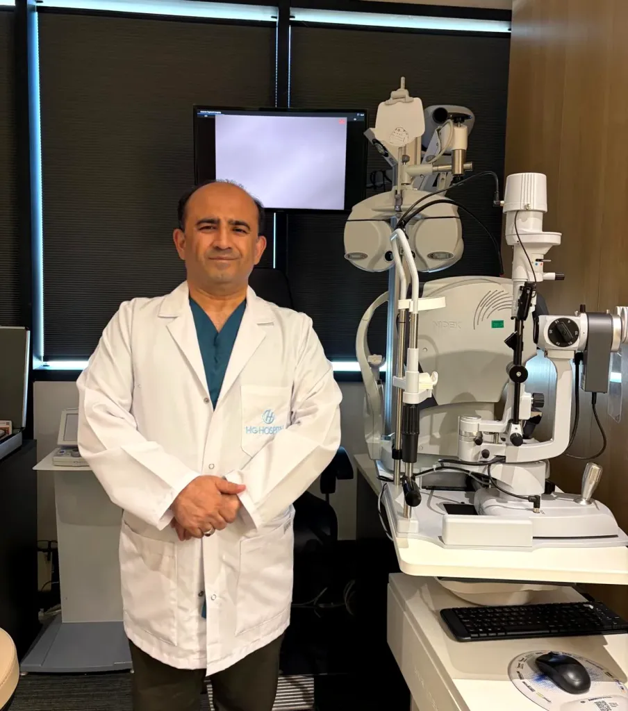
The keratoconus examination, diagnosis and corneal follow-up are carried out by Dr. Nejmi Öztürk, ophthalmologist and eye surgeon, at HG Hospital in Kahramanmaraş, Türkiye. The stage is determined with the corneal map and thickness measurement; progression is documented with measurements taken at intervals, and the treatment step is chosen accordingly.
Your suitability for methods such as cross-linking and combined laser is assessed with the examination and measurements at HG Hospital in Kahramanmaraş; the method that suits you is planned together.
HG Hospital is a private hospital contracted with the Turkish social security institution (SGK); patients with valid SGK coverage may use it, although a private-hospital co-payment may apply to examinations and tests. Cross-linking may be covered by SGK under certain conditions; intracorneal ring segments are not covered. Patients without SGK coverage, such as international patients and visitors from abroad, pay privately or through their private or travel health insurance; the hospital checks coverage before treatment. Fee information is given by the hospital after the examination, and insurance coverage is clarified at the consultation.
Patients from Elbistan, Afşin, Göksun, Ekinözü and Nurhak come to HG Hospital in the center of Kahramanmaraş for the examination; Dr. Öztürk does not see patients in Elbistan. For travel and the same-day test plan, see Elbistan and the surrounding area.
Keratoconus screening and planning with RLES AI
In the assessment of keratoconus, Op. Dr. Nejmi Öztürk uses RLES AI, an explainable artificial intelligence (XAI)-based clinical decision support system, as a second layer of analysis. The system does not diagnose; it independently re-reads the corneal measurements and flags findings that could be overlooked.
- Early-stage screening: Signs of ectasia in the topography and thickness maps are evaluated.
- Contact-lens-related misreading: An attempt is made to distinguish the temporary corneal shape distortion caused by lens wear from true keratoconus.
- Treatment planning: Thickness and safety measures are calculated for options such as cross-linking (CXL) and intracorneal rings.
- Follow-up: Progression is monitored by comparing successive measurements.
This assessment does not replace the physician’s decision; your physician makes the treatment decision and the plan together with the examination findings. RLES AI is a clinical decision support application and is intended solely for use by physicians.
Dr. Nejmi Öztürk sees patients at HG Hospital in Kahramanmaraş, Türkiye. For patients travelling from other districts or from abroad, examination, tests and treatment planning can be arranged for the same day.
Hospital switchboard (within Türkiye): 444 46 46 · E-mail: info@nejmiozturk.com · HG Hospital, Üngüt Mevkii, Prof. Dr. Necmettin Erbakan Blv. No:209, Onikişubat / Kahramanmaraş, Türkiye
Frequently asked questions
Does keratoconus progress, or does it stop by itself?
Keratoconus is a progressive disease; progression is fast particularly in adolescence and the 20s. After the age of 35–40, progression slows down or stops in most patients because the cornea stiffens naturally. This does not apply to every patient; whether it is progressing can be known only with regular corneal measurements.
My glasses prescription keeps increasing; could it be keratoconus?
A constantly increasing prescription, particularly an increase in astigmatism, is the most common warning sign of keratoconus. However, not every increase in prescription is keratoconus; myopia can also increase during youth. Corneal topography and tomography make the distinction; these measurements take a short time and do not touch the eye.
Is keratoconus genetic? Will my child inherit it?
There is a genetic predisposition in keratoconus, but the disease is not passed on by simple inheritance. Some patients have another family member with keratoconus. Screening the children and siblings of a person with keratoconus with a corneal map during adolescence is recommended.
Does rubbing the eyes cause keratoconus?
Eye rubbing is the most important modifiable risk factor associated with the onset and progression of keratoconus. Repeated vigorous rubbing applies mechanical pressure to the cornea. People with allergies are advised to have the itching treated and to give up the habit of rubbing.
Which lens is used in keratoconus? Are glasses enough?
At an early stage glasses may be enough. As the astigmatism becomes irregular, rigid gas-permeable, hybrid or scleral lenses come into consideration. The physician decides which lens is suitable using the corneal map and a trial fitting; a lens corrects vision but does not halt progression.
Is there an operation for keratoconus? When is it needed?
The interventional methods in keratoconus are cross-linking, intracorneal ring segments and corneal transplant. Cross-linking is used to halt progression, ring segments to reduce irregular astigmatism at a moderate stage, and a transplant at an advanced stage when vision cannot be achieved with lenses. Which method is needed is determined by the stage, the thickness and the state of progression.
Can someone with keratoconus have laser eye surgery?
Standard refractive laser operations (such as LASIK and SMILE) are not performed on an eye with keratoconus; the cornea can weaken further. In suitably selected patients, only a topography-guided surface laser that removes a limited amount of tissue can be applied, together with cross-linking. This method aims to reduce the irregularity, not to remove the need for glasses.
My eyes itch a lot from the dust after the earthquake; does this increase the risk of keratoconus?
Dust and allergens increase itching, and itching increases eye rubbing; rubbing is a known risk factor for keratoconus. It would not be correct to say that dust itself causes keratoconus; however, treating the itching and avoiding rubbing are important. A corneal map is recommended for people whose itching continues and whose prescription is changing.
How much does keratoconus treatment cost?
Fee information is given by the hospital after the examination; insurance coverage is clarified at the consultation. Cross-linking may be covered by SGK under certain conditions; intracorneal ring segments are not covered. A private-hospital co-payment may apply at HG Hospital.
I live in Elbistan; where should I go for a keratoconus examination?
Dr. Öztürk sees his patients only at HG Hospital in the center of Kahramanmaraş; he does not examine patients in Elbistan. The road from Elbistan to Kahramanmaraş is about 148 km and takes 2 hours. The examination, corneal measurements and treatment planning are completed on the same day as far as possible; you are advised to bring your old glasses prescriptions and measurement printouts.
Sources
- Turkish Ophthalmological Society — todnet.org
- American Academy of Ophthalmology (AAO) — aao.org
- European Society of Cataract and Refractive Surgeons (ESCRS) — escrs.org
- National Eye Institute (NEI) — nei.nih.gov
Related pages
- Corneal cross-linking (CXL)
- Combined cross-linking and topography-guided laser
- Corneal diseases and corneal transplant
- Laser eye surgery and suitability screening
This page is for information only; diagnosis and treatment decisions are made after an examination. The results of any surgical or interventional procedure may vary from person to person. You are advised to obtain detailed advice from your physician before any procedure.

Ophthalmologist and eye surgeon. Graduate of Erciyes University Faculty of Medicine; completed his residency at Ankara Atatürk Training and Research Hospital. With 21 years of experience in cataract and refractive surgery, strabismus, glaucoma, retina and oculoplastic surgery, he sees patients at HG Hospital in Kahramanmaraş, Türkiye. Biography and training
Dr. Nejmi Öztürk
Ophthalmologist and eye surgeon
HG Hospital, Üngüt Mevkii, Prof. Dr. Necmettin Erbakan Blv. No:209, Onikişubat / Kahramanmaraş
+90 506 377 47 68WhatsAppAppointment formHospital switchboard (within Türkiye): 444 46 46 · info@nejmiozturk.com
- Private patients and private health insurance are welcome; patients with SGK coverage may use it (conditions are confirmed by the hospital)
- Same-day examination and test planning for patients travelling from other districts or from abroad
- Getting here and patient information
- Patients from Elbistan and the region