
Eyelid Aesthetic Surgery (Blepharoplasty)
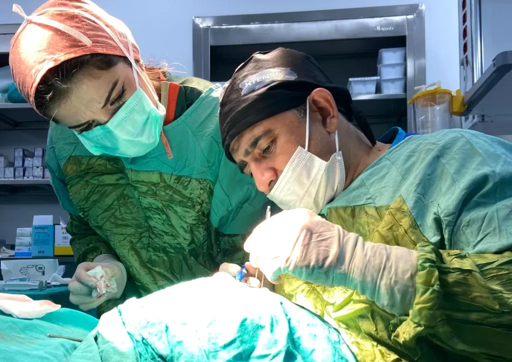
At HG Hospital in Kahramanmaraş, Türkiye, by Dr. Nejmi Öztürk, ophthalmologist — upper eyelid surgery, lower eyelid surgery and ptosis (droopy eyelid) surgery
- What eyelid aesthetic surgery is — the anatomy of the lid, what is checked at the examination, the functional versus cosmetic distinction
- Upper eyelid surgery — excess skin on the upper lid (dermatochalasis), how it is done, recovery and risks
- Lower eyelid surgery — bags in the lower lid, the sub-lash and inner-surface approaches, recovery and risks
- Ptosis (droopy eyelid) — a separate condition: its causes, why it matters in children, levator surgery and the frontalis sling
What is eyelid aesthetic surgery?
Eyelid aesthetic surgery (blepharoplasty) is the reshaping of excess skin, loose muscle and forward-bulging fat tissue in the upper and/or lower eyelid. The aim is to reduce the tired, full look around the eyes without disturbing the structure that allows the lid to open and close comfortably.
The eyelid is made up of thin skin, a layer of muscle beneath it, the cartilage-like tarsal tissue that gives the lid its shape, and the membranes that separate these layers. With age the skin loses its elasticity, the membrane that holds the fat pads inside the lid loosens, and the fat becomes more prominent at the front. In the upper lid this shows itself as a fold of skin hanging over the lashes, and in the lower lid as bags.
Eyelid aesthetic surgery and ptosis (a droopy eyelid) are two different conditions, and they are covered under separate headings on this page. In short: in blepharoplasty the problem is excess skin and fat, whereas in ptosis it is weakness of the muscle that lifts the lid. The two can also be present together in the same person; how much each one contributes is determined by the measurements taken at the examination.
What is checked at the examination?
- Eyelid measurements: The distance from the upper lid edge to the pupil, how far the lid opens and the symmetry between the two eyes are measured.
- Brow position: A drooping brow can make the excess lid skin look greater than it is. If the appearance improves noticeably when the brow is gently lifted with a finger, the real problem may be in the brow.
- Tear and corneal assessment: The amount of tears and the surface of the cornea are examined; signs of dry eye change both the surgical plan and the aftercare.
- Lower lid tightness: The looseness of the lower lid is assessed; in a loose lid, removing skin alone can cause the lid to turn outward.
- Visual field test: If the sagging upper lid restricts vision, a visual field test is requested in order to document this.
- General health: Thyroid disease, the use of blood thinners, smoking and any condition that affects wound healing are asked about.
Functional or cosmetic?
This distinction matters both for the treatment plan and for payment. If the sagging of the upper lid narrows the visual field, pushes the lashes into the eye or causes headaches because of constant brow raising, the situation is considered functional; in these cases an assessment within SGK (Turkish social security) coverage is possible with a visual field test and photographic documentation. Procedures aimed only at improving appearance are considered cosmetic and fall outside insurance coverage. Which group you belong to becomes clear after the examination and the tests; the coverage conditions are confirmed by the hospital before the procedure.
Upper eyelid surgery (upper blepharoplasty)
Upper eyelid surgery is the removal of the excess skin that has built up in the upper lid and, when needed, of fat tissue. In medicine this excess skin on the lid is called dermatochalasis. When the skin hangs over the lashes the eye looks “closed” or tired; in advanced cases the visual field narrows when looking upward, and the person tries to see by raising the eyebrows without being aware of it.
Who is it suitable for?
- People with a fold of skin that reaches the lash line or covers the lashes on the upper lid
- People who notice that their visual field narrows when looking upward, or who feel a heaviness in the lids towards the end of the day
- People who describe tiredness in the forehead and around the brows because of constant brow raising
- People who find it difficult to apply eye make-up because the lid crease has disappeared
- People whose general health is suitable for surgery and whose expectations are realistic
In people with severe signs of dry eye, uncontrolled thyroid eye disease, a condition that would impair wound healing, or recent eye surgery, the procedure is postponed or the plan is changed. The use of blood thinners is adjusted before surgery with the approval of the doctor who prescribed them.
How is it done?
- Marking: The amount of skin to be removed is marked along the natural lid crease while the patient is sitting up. A certain margin of skin is always left so that the lid can still close comfortably.
- Anesthesia: A local anesthetic is applied to the lid; there is a burning sensation for a few seconds during the injection, after which the area goes numb. Sensations of touch and pulling during the procedure are normal. Light intravenous sedation can be added on request.
- Incision: The incision is made within the natural lid crease, so that the mark stays hidden inside the crease when the eye is open.
- Correction: The excess skin is removed; if needed, a thin strip of the lid muscle and the fat tissue that has become prominent at the front are reshaped. The fat tissue is not removed entirely — a measured approach is taken so that the area around the eye does not look hollow.
- Closure: The skin is closed with very fine stitches. If non-dissolving stitches have been used, they are removed at the clinic after 5–7 days.
- Discharge: The eyes are not covered; cold compresses and an antibiotic ointment are recommended. The patient goes home the same day with a companion.
Recovery
| Period | What to expect | What to do |
|---|---|---|
| First 48 hours | Swelling and bruising are at their most noticeable | Cold compresses, sleeping with the head raised, avoiding straining |
| Days 3–7 | The swelling begins to subside and the bruising changes color | Using the ointment, keeping the stitches dry, no make-up |
| Weeks 1–2 | The stitches are removed; a return to desk work is usually possible | Staying away from heavy exercise and work that involves bending over |
| Weeks 3–6 | The incision line may be pink and feel slightly firm | Sun protection, the scar care your doctor recommends |
| Months 3–6 | The line fades and the swelling settles completely | Follow-up examination |
Risks and limitations
- Temporary dryness and stinging: Common in the first weeks after surgery; managed with artificial tears. The complaint can be more noticeable in people who had dry eye beforehand.
- A difference between the two sides: The swelling may resolve at different rates; if a lasting asymmetry remains, a further correction may be needed.
- Incomplete closure of the lid: This can occur if too much skin is removed, which is why a measured approach is taken when marking.
- Scarring: A fine line remains in the lid crease. How noticeable it is depends on the person’s skin type.
- Bleeding, infection: Rare; following the instructions reduces how often they occur.
- What to expect from the result: The procedure reshapes the excess skin and fat on the lid; on its own it does not remove crow’s feet, a drooping brow or bruised-looking shadows on the lower lid.
Lower eyelid surgery (lower blepharoplasty)
Lower eyelid surgery is the repositioning or partial removal of the fat tissue that forms bags in the lower lid and, when needed, the support of a lid that has become loose. In the lower lid the main problem is most often not excess skin but the loosening of the membrane that holds the fat pads, which lets the fat become prominent at the front; for this reason the approach differs from that used in the upper lid.
Who is it suitable for?
- People with persistent bags in the lower lid that do not change between morning and evening
- People who describe shadowing under the eye because of prominent fat
- People with looseness and fine wrinkling of the lower lid skin
- People whose general health is suitable and whose expectations are realistic
Swelling that is noticeable in the morning and decreases during the day is usually due to fluid retention, an allergy or a kidney or thyroid-related cause and does not require surgery; in that case the underlying cause is investigated first. In people with marked lower lid looseness, removing skin alone can cause the lid to turn outward, so an additional step that tightens the lid is planned.
How is it done?
| Approach | Who it is preferred for | How it is done |
|---|---|---|
| From the inner surface (transconjunctival) | People with little excess skin, whose main problem is fat bags | The lid is entered from its inner surface; the fat is repositioned or partly removed. Because no external incision is made, it leaves no visible mark. |
| Under the lashes (transcutaneous) | People whose fat bags are accompanied by clearly excess skin | Through a fine incision just under the lashes, the fat is reshaped and the excess skin is removed. |
| Combined with lid tightening | People found to have lower lid looseness | In addition to one of the approaches above, the lid is supported at its outer corner, which prevents it from being pulled downward. |
The procedure is usually performed under local anesthesia, with light intravenous sedation if requested. Which approach is suitable is decided at the examination, by assessing the amount of excess skin, the degree of fat bagging and the tightness of the lower lid.
Recovery
- First 48–72 hours: Swelling and bruising in the lower lid can be more noticeable than in the upper lid; because of gravity, the bruising may travel down towards the cheek. Cold compresses and sleeping with the head raised are recommended.
- Week 1: If a sub-lash incision was made, the stitches are removed during this period. Procedures done from the inner surface usually leave no stitches to remove.
- Weeks 2–3: The bruising largely resolves and becomes coverable with make-up. Heavy exercise is restarted gradually after this period.
- Months 1–3: A feeling of mild tightness and firmness in the lower lid may continue; it softens with time.
- Months 3–6: The final appearance settles and a follow-up examination is carried out.
Risks and limitations
- The lid being pulled downward (retraction) or turning outward (ectropion): This can occur particularly if lid looseness is overlooked, which is why assessing tightness is an inseparable part of the plan.
- Prolonged swelling: Lower lid swelling can take longer to resolve than in the upper lid.
- Dryness and watering: May occur in the first weeks; managed with artificial tears.
- Scarring: A fine line remains with a sub-lash incision; a procedure done from the inner surface leaves no external mark.
- What to expect from the result: The procedure reduces the bagging; on its own it does not remove dark coloring under the eye, the vascular shadowing beneath the skin or expression lines.
Ptosis (droopy eyelid)
Ptosis is an upper eyelid that sits lower than it should. Normally the upper lid covers the top 1–2 mm of the colored part of the eye (the iris). When the lid drops further, it can cover part or all of the pupil. This is a separate condition from eyelid aesthetic surgery: the problem is not excess skin but the weakening of the muscle that lifts the lid (the levator muscle) or of the membrane that connects this muscle to the lid (the levator aponeurosis). Its treatment is different too — removing skin alone does not correct ptosis.
What are the causes?
- Age-related (aponeurotic) ptosis: The most common type. The levator aponeurosis loosens with aging, long-term contact lens wear, eye rubbing or after previous eye surgery.
- Congenital ptosis (present from birth): The levator muscle has not developed sufficiently; it is noticed from infancy and can affect one or both eyes.
- Nerve-related (neurogenic) ptosis: Seen when the nerve that stimulates the lifting muscle is damaged, or in conditions such as Horner syndrome.
- Muscle-related (myogenic) ptosis: Seen in muscle diseases such as myasthenia gravis; it may get worse and better during the day.
- Mechanical ptosis: A lump, swelling or scar tissue on the lid pulls the lid down.
A droopy lid that has just started, is progressing rapidly, or comes with double vision or a difference in pupil size requires urgent assessment; this picture can be a sign of an underlying problem in the nervous system.
Why does ptosis matter in children?
In childhood a droopy lid can block the development of vision, either by covering the pupil or by pressing on the cornea and creating astigmatism; this leads to lazy eye (amblyopia). For this reason, ptosis in children is not a matter of appearance but a matter of visual development. In severe cases where the pupil is covered, surgery is planned at an early stage; in mild cases it is possible to wait until the preschool years with regular vision follow-up. The details are on the strabismus and lazy eye page.
How is it operated on?
| Method | Who it is preferred for | How it is done |
|---|---|---|
| Levator surgery (aponeurosis advancement / resection) | Age-related and congenital ptosis with good or moderate levator function | Through an incision in the lid crease, the membrane that connects the muscle to the lid is shortened and stitched back to the lid cartilage (tarsus); the lid is set at the desired level. |
| Müller muscle surgery (approach from the inner surface) | Mild ptosis; people who respond well to the drop test | The lid is entered from its inner surface and the muscle is shortened without leaving an external mark. |
| Frontalis sling | Congenital ptosis with weak levator function; some nerve- and muscle-related cases | The lid is connected to the forehead muscle with a silicone sling or with a strip of the patient’s own thigh tendon (fascia lata); the patient opens the lid by raising the brow. |
In adults the procedure is usually done under local anesthesia. The reason is that the patient can be asked to open and close the eye during the operation, so that the lid height can be fine-tuned at that moment. General anesthesia is preferred in children and in cases that require a frontalis sling. If the droopy lid is also accompanied by clearly excess skin, ptosis surgery and upper eyelid surgery can be planned in the same session; this decision is made according to the measurements.
Recovery
- First 48 hours: Swelling and bruising are noticeable; cold compresses are applied and the head is kept raised.
- Week 1: The stitches are removed. During this period the lid may look higher or lower than it should; the result is not assessed until the swelling has resolved.
- Weeks 2–4: The lid level begins to settle. Because the lid may not close completely in the first weeks, a night-time ointment may be recommended.
- Months 2–3: The lid height and symmetry become clear; the result is assessed during this period.
- Revision: In a proportion of patients the lid may end up above or below the intended level and a second correction may be needed. This is a known limitation of the surgery and is discussed before the operation.
Risks and limitations
- Under-correction or over-correction: The lid may not settle exactly at the intended level; a revision may be needed.
- Asymmetry between the two sides: May be seen particularly in one-sided cases.
- Incomplete closure of the lid (lagophthalmos): May be seen in the early period because the lid has been raised; drops and ointment are used to protect the cornea. This can be more marked after a frontalis sling.
- An increase in dry eye complaints: Expected in people who had dry eye beforehand; managed with treatment.
- Irregularity of the lid crease and, rarely, bleeding and infection.
What changes in the eyelid?
A diagram showing that a droopy eyelid and excess skin are separate problems.
- Droopy eyelidWhen the muscle that lifts the upper lid weakens, the lid drops over the pupil. The upper part of the visual field may be covered; the person tries to see by raising the eyebrow or tilting the head back.
- Excess skinWith age, excess skin that builds up on the upper lid can limit vision. This is different from muscle weakness; the two can also exist together.
Eyelid aesthetic surgery in Kahramanmaraş
Upper eyelid surgery, lower eyelid surgery and ptosis surgery are performed at Private HG Hospital in Kahramanmaraş by Dr. Nejmi Öztürk, ophthalmologist. The examination, the eyelid measurements, a visual field test where needed and the operation are all planned at the same hospital. For patients coming from Elbistan, Afşin, Göksun, Ekinözü and Nurhak, the examination and the tests are scheduled on the same day as far as possible; the operation is planned for a separate day because it requires preparation. To make an appointment you can use the details on the contact page.
Inflammation of the lid margin (blepharitis) can cause swelling that mimics sagging; in that case the inflammation is treated first. Watering is covered on the blocked tear duct page, and dryness after surgery on the dry eye treatment page.
Dr. Nejmi Öztürk sees patients at HG Hospital in Kahramanmaraş, Türkiye. For patients travelling from other districts or from abroad, examination, tests and treatment planning can be arranged for the same day.
Hospital switchboard (within Türkiye): 444 46 46 · E-mail: info@nejmiozturk.com · HG Hospital, Üngüt Mevkii, Prof. Dr. Necmettin Erbakan Blv. No:209, Onikişubat / Kahramanmaraş, Türkiye
Frequently asked questions
Are eyelid aesthetic surgery and droopy eyelid surgery the same thing?
No. In eyelid aesthetic surgery (blepharoplasty) the excess skin and fat are reshaped; in a droopy eyelid (ptosis) the muscle that lifts the lid is weak, and the surgery works on that muscle. The two can be present together in the same person and can be done in the same session when needed, but they are different procedures.
Should I have upper or lower eyelid surgery?
It depends on your complaint. Skin hanging over the lashes and a narrowing visual field concern the upper lid, while persistent bags in the lower lid concern the lower one. The two can also be planned together; the decision is made according to the measurements taken at the examination.
Is pain felt during the procedure?
A burning sensation is felt for a few seconds during the injection of the local anesthetic; after that the area goes numb. Sensations of touch, pulling and pressure throughout the procedure are normal. If you feel uncomfortable, more anesthetic is given, and light intravenous sedation can be added on request.
When can I go back to work?
Most patients return to desk work within 1 week; it can take 2–3 weeks until the bruising becomes coverable with make-up. For heavy physical work and sport, follow the period your doctor gives you.
Will there be a scar from the stitches?
In the upper lid the incision stays inside the natural lid crease and fades with time. In the lower lid, procedures done from the inner surface leave no external mark; a sub-lash incision leaves a fine line. How noticeable the mark is depends on the person’s skin type.
How can I tell whether it is a drooping brow or an eyelid problem?
If the appearance improves noticeably when you gently lift the brow with a finger in front of a mirror, the problem is most likely in the brow. If the lid edge stays close to the pupil even with the brow raised, ptosis is considered. The distinction becomes clear with the lid and brow measurements taken at the examination.
Does SGK cover it?
If the sagging of the upper lid narrows the visual field in a way that can be documented, the procedure is considered functional and may be assessed within SGK (Turkish social security) coverage after a visual field test and photographic documentation. Procedures aimed only at appearance are considered cosmetic and fall outside coverage. The coverage conditions are confirmed by the hospital before the procedure.
Is the result permanent?
The skin that has been removed does not come back, but the aging process continues; over the years the lid and brow tissue may loosen again. In ptosis surgery, the lid level may change to some degree over time in a proportion of patients.
Where is eyelid aesthetic surgery done in Kahramanmaraş?
Dr. Nejmi Öztürk performs eyelid aesthetic surgery and ptosis operations at Private HG Hospital in Onikişubat. The examination, the measurements and the operation are planned at the same hospital. Appointment details are on the contact page.
I am coming from Elbistan; can the examination and the operation be on the same day?
Usually the examination, the eyelid measurements and, if needed, a visual field test are done first, and the operation is planned for a separate day, because adjusting blood thinners and the documentation process take time. For patients coming from far away, stitch removal and check-up days are combined as far as possible.
Sources
- Turkish Ophthalmological Society — todnet.org
- American Academy of Ophthalmology (AAO) — aao.org
- National Eye Institute (NEI) — nei.nih.gov
Related pages
- Strabismus and lazy eye
- Blocked tear duct
- Dry eye treatment
- Blepharitis (inflammation of the lash line)
This page is for information only; diagnosis and treatment decisions are made after an examination. The results of any surgical or interventional procedure may vary from person to person. You are advised to obtain detailed advice from your physician before any procedure.

Ophthalmologist and eye surgeon. Graduate of Erciyes University Faculty of Medicine; completed his residency at Ankara Atatürk Training and Research Hospital. With experience in ophthalmology since 2005 in cataract and refractive surgery, strabismus, glaucoma, retina and oculoplastic surgery, he sees patients at HG Hospital in Kahramanmaraş, Türkiye. Biography and training
Dr. Nejmi Öztürk
Ophthalmologist and eye surgeon
HG Hospital, Üngüt Mevkii, Prof. Dr. Necmettin Erbakan Blv. No:209, Onikişubat / Kahramanmaraş
+90 506 377 47 68WhatsAppAppointment formHospital switchboard (within Türkiye): 444 46 46 · info@nejmiozturk.com
- Private patients and private health insurance are welcome; patients with SGK coverage may use it (conditions are confirmed by the hospital)
- Same-day examination and test planning for patients travelling from other districts or from abroad
- Getting here and patient information
- Patients from Elbistan and the region