
Cataract Surgery
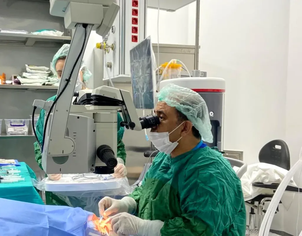
Performed by Dr. Nejmi Öztürk, ophthalmologist and eye surgeon, at HG Hospital in Kahramanmaraş, Türkiye — with monofocal and toric lens options
- A cataract is a clouding of the eye’s lens; it cannot be reversed with drops or glasses, and its treatment is surgery.
- The surgery is done with anesthetic drops, through a 2–3 mm incision, using the ultrasound (phacoemulsification) method, in 15–30 minutes; you go home the same day.
- For patients with valid SGK coverage, the standard monofocal lens is covered; with toric, EDOF and trifocal lenses the lens surcharge is paid by the patient.
- For patients coming from Elbistan and the surrounding districts, the day of surgery and the day 1 check-up are planned together.
What is a cataract?
A cataract is a loss of transparency and clouding of the natural lens inside the eye. The eye’s lens is a clear structure, similar to a camera lens, that focuses light onto the retina (the light-sensitive layer at the back of the eye). With age the proteins of the lens change; the lens yellows and becomes dull. Light no longer reaches the retina sharply, and vision becomes hazy and faded.
The most common cause is aging; after the age of 60, many people have some degree of cataract. Diabetes, long-term use of cortisone (steroids), eye injuries, inflammation inside the eye, smoking and intense sunlight can speed up cataract formation. Rarely, babies are born with a cataract (congenital cataract); this requires early diagnosis and treatment.
A cataract is not contagious and cannot be reversed with medication, drops or glasses. The treatment is to remove the clouded lens surgically and replace it with an artificial intraocular lens.
What are the symptoms of a cataract?
The most common symptom of a cataract is gradually increasing blurred vision that glasses do not correct. The symptoms progress over months, sometimes years; they may not be noticed at the start.
- Blurred, hazy or foggy vision; a feeling of looking through a dirty window
- Fading and yellowing of colors; white looking cream-colored
- Glare and scattering of light around headlights when driving at night, sensitivity to light
- Frequent changes in glasses prescription; new glasses becoming inadequate within a short time
- Double or shadowed vision when looking with one eye
- Needing more light than before when reading
- A temporary improvement in near vision (the feeling “I no longer need my reading glasses”); this is caused by a change in the power of the lens and is not permanent
These symptoms can also be seen in other diseases such as macular degeneration, glaucoma (high eye pressure) and retinal damage due to diabetes. For this reason, the diagnosis is made with an eye examination.
When should a cataract be operated on?
A cataract is operated on when the loss of vision starts to make your daily life difficult. If driving, reading, watching television, working or recognizing faces is becoming harder and a change of glasses is not enough, the time for surgery has come. There is no need to wait for the cataract to “ripen”; on the contrary, very advanced and hardened cataracts make the surgery more difficult and can lead to problems with eye pressure and inflammation.
In some situations surgery may be recommended even though the cataract does not greatly impair vision: examples are a cataract that prevents examination of the retina (particularly in people with diabetes), a cataract that raises eye pressure, and a cataract that creates a large difference in prescription between the two eyes.
Who is it not suitable for?
Cataract surgery is postponed in patients with an active eye infection or uncontrolled inflammation inside the eye until these problems have been treated. Patients whose blood sugar or blood pressure is poorly controlled are asked to have these values brought under control first. Macular degeneration, advanced glaucoma or retinal damage do not rule out surgery, but the gain in vision may be limited, and this is discussed openly with you before the surgery. For an early cataract that does not affect daily life, regular follow-up is recommended instead of surgery.
Which examinations and tests are done before surgery?
The decision for surgery and the choice of lens are made with a detailed eye examination and biometry measurements. At the examination your visual acuity and glasses prescription are measured; the density of the cataract is assessed with the slit lamp (eye microscope); eye pressure is measured; and the retina and macula are examined with the pupil dilated with drops.
- Optical biometry: The length of the eye and the curvature of the cornea (the clear front layer) are measured; the power of the lens to be implanted is calculated from these measurements.
- Corneal topography: The amount and pattern of astigmatism (the cornea being more curved in one direction) are determined; this measurement decides whether a toric lens is needed.
- OCT (optical coherence tomography of the eye): The macula and the layers of the retina are examined; diseases that could limit vision are detected in advance.
- Endothelial cell count: The number of cells on the inner surface of the cornea is measured in patients where this is considered necessary.
- Eye ultrasound: If the cataract is dense enough to prevent a view of the retina, the back of the eye is examined with ultrasound.
Be sure to tell us about the medicines you take, particularly blood thinners and prostate medicines; some prostate medicines affect the behavior of the pupil during surgery. Most blood thinners are not stopped for cataract surgery done with anesthetic drops; the decision is made together with the doctor who prescribed the medicine. In people with diabetes, blood sugar is expected to be well controlled before surgery.
How is cataract surgery performed?
Today, cataract surgery is done with the phacoemulsification method, in which the cloudy lens is broken up with ultrasound waves and removed through a small incision. The procedure is usually done with anesthetic drops; stitches are usually not needed. The steps are as follows:
- Preparation: Drops that dilate your pupil and numb the eye are applied; the area around the eye is cleaned with an antiseptic solution.
- Anesthesia: In most patients anesthetic drops alone (topical anesthesia) are sufficient; if necessary, additional numbing is given around the eye with a small injection. General anesthesia is preferred for children and for patients who would not be able to cooperate during the surgery.
- Incision: A small incision about 2–3 mm wide is made at the edge of the cornea. This incision is designed to seal by itself.
- Opening the capsule: A round opening is made in the front surface of the thin membrane (capsule) that surrounds the lens (capsulorhexis).
- Breaking up the lens: The hard core of the lens is broken up with a fine probe that delivers ultrasound energy and is removed by suction through the same probe. The remaining soft parts are cleaned away; the posterior capsule is left in place.
- Implanting the artificial lens: The foldable intraocular lens is delivered through the same incision with a thin injector and unfolds inside the capsular bag, settling into position. If a toric lens is used, it is rotated to the axis calculated beforehand.
- Finishing: The incision is checked for leaks, an antibiotic may be given inside the eye, and a protective shield is placed over the eye.
The surgery usually takes 15–30 minutes; with preparation and rest, the time spent at the hospital comes to a few hours. During the procedure you feel light, mild pressure and touch; after the surgery there may be stinging, watering and a gritty feeling for a few hours. If both eyes have a cataract, the eyes are usually operated on separate days.
Cataract surgery step by step
Removal of the clouded natural lens and placement of an intraocular lens, shown schematically on a cross-section of the front of the eye.
- Small incisionAfter numbing eye drops, an incision of about 2–3 mm is made at the edge of the cornea; in most cases no stitches are needed.
- Opening the front capsuleA circular opening is made in the front of the capsule that surrounds the lens; the rest of the capsule is preserved as the bed for the new lens.
- Breaking up and removing the cataractThe clouded lens is broken up with ultrasound energy and removed by suction through the same incision.
- Placing the intraocular lensThe chosen lens is inserted in folded form, unfolds inside the capsular bag and stays centred. The procedure usually takes 15–20 minutes.
What is a monofocal lens?
A monofocal intraocular lens is the standard artificial lens, which focuses light at a single distance. It is usually chosen so that distance vision is clear; glasses are needed for near tasks such as reading, sewing and using a phone. It has been used for many years, its results are well known, and it is the most commonly chosen lens type in cataract surgery.
A monofocal lens can also be used in patients who have an additional eye disease besides the cataract (macular degeneration, advanced glaucoma), because it does not split light and therefore does not cause a loss of contrast (the ability to distinguish shades). Complaints of night halos and glare are fewer than with other lens types.
Depending on preference and the structure of the eye, one eye can be set for distance and the other slightly for near (monovision). This arrangement reduces the need for glasses to some extent, but not everyone can adapt to it; suitability is assessed at the examination. For patients with valid SGK coverage, the standard monofocal lens is covered.
What is a toric lens?
A toric intraocular lens is a lens that also corrects astigmatism during cataract surgery. In astigmatism the cornea is more curved in one direction; light is focused on the retina as a line rather than a point, and the image is blurred or shadowed at every distance. A standard lens does not correct astigmatism; in a patient with significant astigmatism, glasses are still needed for distance after surgery.
A toric lens carries additional power along a particular axis. Before surgery, the degree and direction of the astigmatism are measured with corneal topography and biometry, and the axis at which the lens will be placed inside the eye is calculated. During surgery this axis is marked on the cornea (by hand or with image-guided systems), and the lens is rotated into position according to this mark.
A toric lens is usually considered in patients with regular astigmatism of one diopter or more; in irregular astigmatism such as keratoconus, the expected benefit is more limited. Rarely, the lens may rotate off its axis in the first days; in that case it is repositioned with a small additional procedure. A toric lens can be monofocal, or it can have a trifocal or EDOF design. If a toric lens is chosen, a surcharge may apply; coverage conditions are clarified at the examination.
What lens options are there?
In cataract surgery there are four main lens groups: monofocal, toric, EDOF and trifocal (multifocal, known locally as the “smart lens”). The choice is made together according to the health of your eye, the amount of astigmatism, your occupation, whether you drive at night and how far you want to be free of glasses.
| Lens type | Clear vision distance | Astigmatism correction | Night halos and glare | Need for glasses | SGK coverage |
|---|---|---|---|---|---|
| Monofocal | Distance (or near if preferred) | No | Low | Needed for near | Standard lens is covered |
| Toric monofocal | Distance; astigmatism corrected | Yes | Low | Needed for near | A surcharge may apply |
| EDOF | Distance and intermediate; glasses may be needed for small print | Toric option available | Low–moderate | Occasionally | Lens surcharge paid by the patient |
| Trifocal (multifocal) | Distance, intermediate and near | Toric option available | Moderate; decreases over time | Usually not needed | Lens surcharge paid by the patient |
For detailed information about multifocal lenses, see the trifocal lens (multifocal intraocular lens) and EDOF lens pages. For young patients with a high prescription and no cataract, the ICL (phakic intraocular lens), which preserves the natural lens, is a separate option. A general comparison of all lens options is on the lens surgery page.
Which lens changes what?
The effect of monofocal, toric and multifocal lenses on vision.
- Monofocal lensFocuses on a single distance; when distance vision is planned to be clear, reading glasses are used for near. It is the standard lens type, and coverage by a country’s social security institution is clarified at the consultation.
- Toric lensAlso corrects astigmatism of the cornea. The lens must be aligned along a specific axis, which is marked during the operation.
- Multifocal lensAims at far, intermediate and near vision together. Seeing rings of light around lights at night is more likely than with a monofocal lens.
What is recovery like after surgery?
You go home on the day of cataract surgery; most patients notice a clear improvement in vision from day 1. Full clarity settles within a few weeks; this period varies with the structure of the eye and the type of lens.
Day 1
At the check-up the day after surgery, the protective shield is removed and the eye pressure and the position of the lens are assessed. Vision may be hazy at this stage; this is expected. Antibiotic and anti-inflammatory drops are started; you will be asked to note the times of the drops. Mild redness, stinging and watering are normal.
Week 1
Vision becomes clearer day by day. This week, do not rub your eye, keep water and soap out of it, do not lift heavy objects and limit tasks that involve bending over; you may be asked to sleep with the protective shield at night. Reading, watching television and light walks are allowed. At the week 1 check-up the drop schedule is updated.
Month 1
The drops are tapered and stopped according to your doctor’s plan; in most patients this takes 3–4 weeks. You usually wait a month before swimming pools, the sea and the hammam (Turkish bath). Your glasses prescription is measured once vision has settled, about 3–4 weeks after surgery. At the month 1 check-up, surgery on the other eye and whether any additional correction is needed are discussed.
If you are coming from Elbistan, Afşin, Göksun, Ekinözü or Nurhak, the day of surgery and the day 1 check-up are planned together; the later check-ups are spaced out according to your travel conditions.
What are the risks and limitations of cataract surgery?
Cataract surgery is a frequently performed operation with well-known results; even so, like every operation, it has risks. Serious problems are rare, but it is important that you know they can occur and recognize their signs.
- Infection (endophthalmitis): Rare, but it requires urgent treatment. Increasing pain, redness and reduced vision in the first days are warning signs.
- Posterior capsule opacification (secondary cataract): The membrane in which the lens is placed can become cloudy over months or years; vision becomes hazy again. This is cleared not with surgery but with a YAG laser procedure lasting a few minutes (described below).
- Macular edema (cystoid macular edema): Can cause blurring of vision weeks after surgery; it usually resolves with drop treatment. The risk is higher in people with diabetes and those with a history of uveitis (inflammation inside the eye).
- Corneal edema: Temporary swelling of the cornea in the first days can blur vision; it usually subsides within days. It may last longer in people with a low endothelial cell count.
- Temporary rise in eye pressure: Can be seen in the first days; it is controlled with drops.
- Posterior capsule tear: The membrane holding the lens can tear during surgery; in that case the lens is placed in a different position or in a second session.
- Retinal detachment (separation of the retina): Rare; the risk is slightly higher in highly myopic people. Sudden flashes of light, floaters and a curtain-like shadow require urgent examination.
- Refractive surprise: Despite the calculations, a small glasses prescription may remain after surgery; it is corrected with glasses or, rarely, with an additional procedure.
Limitations: Cataract surgery corrects only the loss of vision caused by the lens. If there is macular degeneration, glaucoma or retinal damage, vision improves only as far as these diseases allow. Surgery does not remove the need for glasses in every patient; with a monofocal lens, reading glasses are needed, and with multifocal lenses, night halos may be seen.
What are secondary cataract and YAG laser?
Secondary cataract is the clouding over time of the posterior capsule that is left in place during surgery; it is not the cataract “coming back”, because the removed lens does not grow back. Its symptoms are haziness, glare and difficulty reading that reappear months or years after surgery. The treatment is YAG laser capsulotomy: the pupil is dilated with drops, and a small opening is created with the laser in the center of the cloudy membrane. The procedure takes a few minutes, does not require an operating room, and vision usually clears within one to two days; drops are used for a short time afterwards.
Cataract surgery in Kahramanmaraş
In Kahramanmaraş, Türkiye, cataract surgery is performed by Dr. Nejmi Öztürk in the Ophthalmology Department of HG Hospital. Dr. Öztürk is an ophthalmologist working in cataract and refractive surgery; the examination, measurements, surgery and check-ups are all done at the same hospital. The hospital’s address is Üngüt Mevkii, Prof. Dr. Necmettin Erbakan Blv. No:209, 46050 Onikişubat / Kahramanmaraş.
Is cataract surgery covered by SGK or insurance?
HG Hospital is a private hospital contracted with the Turkish social security institution (SGK); patients with valid SGK coverage may use it, although a private-hospital co-payment may apply. For these patients, cataract surgery and the standard monofocal intraocular lens fall within SGK coverage; if a toric, EDOF or trifocal lens is chosen, the lens surcharge is paid by the patient. Patients without SGK coverage, such as international patients and visitors from abroad, pay privately or through their private or travel health insurance; the hospital checks coverage before treatment. Coverage conditions and fee information are clarified by the hospital after the examination.
Planning for patients from Elbistan and the surrounding districts
Dr. Öztürk does not see patients in Elbistan; patients from Elbistan, Afşin, Göksun, Ekinözü and Nurhak come to HG Hospital in Kahramanmaraş. Elbistan–Kahramanmaraş is about 148 km (about 2 hours), Afşin about 123 km and Göksun about 78 km. All measurements, including biometry, are completed on the same day as the examination; a separate preparation day is not needed.
The day of surgery and the next day’s check-up are planned together; patients who wish to can stay one night in Kahramanmaraş or come back the next morning. Because you cannot drive after the surgery, a companion must be with you. Travel and planning details are on the patients from Elbistan and the surrounding area page.
Dr. Nejmi Öztürk sees patients at HG Hospital in Kahramanmaraş, Türkiye. For patients travelling from other districts or from abroad, examination, tests and treatment planning can be arranged for the same day.
Hospital switchboard (within Türkiye): 444 46 46 · E-mail: info@nejmiozturk.com · HG Hospital, Üngüt Mevkii, Prof. Dr. Necmettin Erbakan Blv. No:209, Onikişubat / Kahramanmaraş, Türkiye
Frequently asked questions
How many minutes does cataract surgery take?
Cataract surgery with the phacoemulsification method usually takes 15–30 minutes. With preparation, the drops and rest after the surgery, you spend a few hours at the hospital. With hard and advanced cataracts the time may be a little longer.
Is general anesthesia used in cataract surgery, and what does it feel like?
In most patients only anesthetic drops are used; general anesthesia is preferred for children and for patients who would not be able to cooperate during the surgery. During the procedure you feel light, mild pressure and touch. There may be stinging and watering for a few hours after the surgery.
Will I need glasses after cataract surgery?
This depends on the lens chosen. With a monofocal lens, distance vision becomes clear and glasses are needed for reading; a toric lens also corrects astigmatism. With a trifocal (multifocal) lens, most patients need glasses very little or not at all. The permanent glasses prescription is measured at week 3–4, once vision has settled.
Are both eyes operated on the same day?
If both eyes have a cataract, they are usually operated on separate days. The interval is planned between a few days and a few weeks, depending on the healing of the first eye. This allows the lens calculation for the second eye to be reviewed in light of the first eye’s result.
Can a cataract come back?
No; the removed lens does not grow back. However, the posterior capsule in which the lens is placed can become cloudy months or years later; this is called secondary cataract. Vision is restored with a YAG laser procedure lasting a few minutes, and this procedure is usually done only once.
How much does cataract surgery cost, and does insurance cover it?
For patients with valid SGK coverage, cataract surgery and the standard monofocal lens are covered; a private-hospital co-payment may apply. With toric and multifocal lenses the lens surcharge is paid by the patient. Patients without SGK coverage pay privately or through their private or travel health insurance. Fee information is given by the hospital after the examination; insurance coverage is clarified at the consultation.
Where is cataract surgery performed in Kahramanmaraş?
Dr. Nejmi Öztürk performs cataract surgery at HG Hospital in Onikişubat, Kahramanmaraş. The examination, measurements, surgery and check-ups are all carried out at the same hospital. Appointment and contact details are in the contact section on this page.
Is cataract surgery available in Elbistan?
Dr. Öztürk does not see patients in Elbistan; patients from Elbistan and the surrounding districts have their examination and surgery at HG Hospital in Kahramanmaraş. Because the journey takes about 2 hours, the day of surgery and the day 1 check-up are planned together. The later check-ups are spaced out.
I have diabetes; can I have cataract surgery?
Yes; in people with diabetes, cataract can appear at an earlier age, and surgery can be performed. Blood sugar is expected to be well controlled before surgery, and the retina is assessed with OCT for damage due to diabetes. Because the risk of macular edema is slightly higher, check-ups may be more frequent.
Sources
- Turkish Ophthalmological Society — todnet.org
- American Academy of Ophthalmology (AAO) — aao.org
- European Society of Cataract and Refractive Surgeons (ESCRS) — escrs.org
- National Eye Institute (NEI) — nei.nih.gov
Related pages
- Lens surgery — all intraocular lens options
- Trifocal lens (multifocal intraocular lens)
- EDOF (extended depth of focus) lens
- Eye doctor in Kahramanmaraş — examination and appointments
This page is for information only; diagnosis and treatment decisions are made after an examination. The results of any surgical or interventional procedure may vary from person to person. You are advised to obtain detailed advice from your physician before any procedure.

Ophthalmologist and eye surgeon. Graduate of Erciyes University Faculty of Medicine; completed his residency at Ankara Atatürk Training and Research Hospital. With 21 years of experience in cataract and refractive surgery, strabismus, glaucoma, retina and oculoplastic surgery, he sees patients at HG Hospital in Kahramanmaraş, Türkiye. Biography and training
Dr. Nejmi Öztürk
Ophthalmologist and eye surgeon
HG Hospital, Üngüt Mevkii, Prof. Dr. Necmettin Erbakan Blv. No:209, Onikişubat / Kahramanmaraş
+90 506 377 47 68WhatsAppAppointment formHospital switchboard (within Türkiye): 444 46 46 · info@nejmiozturk.com
- Private patients and private health insurance are welcome; patients with SGK coverage may use it (conditions are confirmed by the hospital)
- Same-day examination and test planning for patients travelling from other districts or from abroad
- Getting here and patient information
- Patients from Elbistan and the region