
Corneal Diseases
Diagnosis, treatment and, when needed, referral to advanced centres — with Dr. Nejmi Öztürk, ophthalmologist and eye surgeon, at HG Hospital in Kahramanmaraş, Türkiye
- Keratitis, corneal abrasion, herpes, dystrophy, oedema and keratoconus
- Which symptoms are emergencies; what to do with contact lenses and work injuries
- Diagnostic methods; treatments ranging from drops to CXL and corneal transplantation
- Treatment and referral at HG Hospital in Kahramanmaraş, and insurance coverage
What is the cornea?
The cornea (often described as the clear “window” of the eye) is the transparent, dome-shaped layer at the very front of the eye, like the glass of a watch. It is about half a millimetre thick and provides most of the eye’s focusing power. The outermost layer (the epithelium) renews itself quickly; the innermost layer (the endothelium) carries the pump cells that keep the cornea clear, and it does not regenerate. The cornea has a very dense network of nerves; even a small scratch causes noticeable pain.
Layers of the cornea
A cross-section of the cornea, to help you understand which layer each treatment concerns.
- Corneal cross-sectionThe cornea is clear and has no blood vessels. On the outside is the self-renewing epithelium, in the middle the stroma, which makes up most of the tissue, and on the inside the endothelium, which keeps the cornea clear. Surface lasers and cross-linking act on the stroma, while corneal transplantation targets the layers concerned.
What are the common corneal diseases?
The problems that most often affect the cornea are infection (keratitis), abrasions and foreign bodies, herpes, inherited dystrophies, oedema and keratoconus.
Keratitis and corneal ulcer
Keratitis is inflammation of the cornea; a corneal ulcer is this inflammation deepening with loss of tissue. It can be caused by bacteria, fungi, viruses or amoebae; sleeping in contact lenses, rinsing lenses with tap water and wearing lenses in swimming pools are the main risks. It shows itself with pain, redness, light sensitivity and a white spot on the cornea; because it can progress within hours, it is an emergency.
Corneal abrasion and foreign body
A corneal abrasion is a scraping-off of the epithelium; it happens with branches, fingernails and dust. In farming, construction, welding and grinding work, metal or plant particles can become embedded in the cornea; in plant-related injuries the risk of fungal infection is high. It is common among agricultural and construction workers in Kahramanmaraş and its districts; protective glasses prevent most cases.
Herpes keratitis
The cold sore virus (herpes simplex) causes branching ulcers on the cornea and, hiding in the nerve, can flare up again years later; each recurrence can leave a scar on the cornea. It is treated with antiviral ointment and tablets; patients with frequent recurrences are given long-term low-dose preventive tablets.
Corneal dystrophies and corneal oedema
Dystrophies are inherited diseases that affect a particular layer of the cornea and are seen in both eyes together. The most widely known is Fuchs endothelial dystrophy: the pump cells decrease, the cornea retains water (oedema) and blurred vision develops that is most noticeable in the morning. Oedema can also occur after cataract surgery. In mild oedema, hypertonic saline drops are used; advanced cases need an endothelial transplant.
Keratoconus
Keratoconus is a thinning of the cornea that makes it bulge forward into a cone shape; it begins at a young age, and eye rubbing and allergy speed up its progression. Frequent changes of glasses prescription and increasing astigmatism are warning signs. Corneal cross-linking (CXL) to halt progression, and special contact lenses and intracorneal rings to improve vision, are described on the keratoconus treatment page.
Pterygium and surface damage due to dry eye
A pterygium (surfer’s eye) can advance across the cornea and cause astigmatism and scarring. Dry eye and blepharitis cause punctate damage to the corneal surface; other corneal treatments are not planned until the surface has been treated.
What are the symptoms? Which symptoms are emergencies?
The symptoms common to corneal diseases are pain, stinging, redness, light sensitivity and blurred vision.
- Come the same day: Pain and redness that begin while wearing contact lenses; a white spot on the cornea; a piece of metal, stone or plant getting into the eye; a chemical splash (rinse with plenty of water for 15–20 minutes beforehand); a sudden drop in vision with severe pain.
- Examination within a few days: Recurring cold-sore-like redness of the eye, blurred vision in the morning, frequent changes of glasses prescription, a feeling of sand in the eye that does not go away.
Diagnosis and treatment are suitable for patients of all ages; contact lens wearers, agricultural and construction workers, and people with keratoconus in the family benefit particularly from regular examinations.
Who is it not suitable for?
- Steroid (cortisone) drops: Not suitable in fungal keratitis or during the active ulcer phase of herpes; they deepen the infection. Steroid drops should not be used on a red eye without an examination.
- Contact lenses and bandage lenses: Not suitable during an active infection or for patients who cannot maintain lens hygiene.
- CXL: Not suitable when the cornea has become too thin, when there is a scar in the centre, or when the keratoconus has stopped progressing.
- Corneal transplantation: Not suitable while there is uncontrolled dry eye, an eyelid problem or an active infection.
Which examinations and tests are done?
The diagnosis of corneal diseases begins with a slit-lamp examination and a dye test; measuring devices are added when needed.
- Slit-lamp examination and fluorescein staining: Scratches, ulcers and surface damage become visible with the dye.
- Corneal topography: A map of the curvature of the cornea; the basic test for keratoconus and astigmatism.
- Pachymetry: Measurement of corneal thickness; used in oedema, keratoconus and assessing suitability for laser.
- Endothelial cell count (specular microscopy): The number of pump cells; used in Fuchs dystrophy and in pre-operative assessment.
- Culture: In an ulcer, a sample is taken from the cornea to identify the organism.
How are the treatments carried out?
Depending on the disease, corneal treatment ranges from drops and ointment to bandage lenses, CXL, intracorneal rings and corneal transplantation.
- Drops and ointment: In a bacterial ulcer antibiotic drops are used hourly at the start, antifungals in fungal infection and antivirals in herpes; steroid is added only on the doctor’s decision. Hypertonic saline drops are given for oedema and artificial tears for surface damage.
- Foreign body removal and treatment of abrasions: After anaesthetic drops, the particle and any rust ring are cleaned away under the slit lamp with a fine-tipped instrument; the procedure takes a few minutes and antibiotic ointment is given. What to expect: slight pressure during the procedure, and stinging for 1–2 days afterwards.
- Bandage contact lens: In painful surface damage it covers the cornea and eases healing; it stays in place from a few days to a few weeks under the doctor’s supervision.
- Corneal cross-linking (CXL): In keratoconus, riboflavin (vitamin B2) drops are applied to the cornea and ultraviolet light is used to stiffen the tissue; it aims to halt progression and does not improve vision. Details are on the cross-linking page.
- Intracorneal rings: Thin rings placed in the cornea reduce the cone shape; they do not halt progression.
- Corneal transplantation: A donated clear cornea is stitched in place of the diseased tissue; there are full-thickness transplants, DALK in which only the front layers are replaced, and DMEK/DSAEK in which only the endothelium is replaced. Transplants are performed at tertiary centres with an eye bank; suitable patients are referred to these centres.
What is recovery like?
Recovery time varies from a few days to a year, depending on the disease.
Day 1 – week 1
After a simple abrasion or foreign body, the epithelium closes in 1–3 days. In keratitis the drops are used very frequently at the start and the patient is checked every 1–2 days; a herpes flare-up settles in 1–2 weeks.
Month 1
As a corneal ulcer heals, the frequency of the drops is reduced; the effect of any remaining scar on vision becomes clear in this period. After CXL the epithelium closes in 3–5 days and vision may fluctuate for 1–3 months.
Long term
Keratoconus and Fuchs dystrophy require long-term follow-up; topography and endothelial cell counts are repeated every 6–12 months. After a corneal transplant, vision settles in 6–12 months. Check-ups for patients from the outlying districts are grouped on the same day; in an emergency, go straight to the nearest emergency department.
What are the risks and limitations?
The most important risk in corneal diseases is that infections and injuries treated late leave permanent scars.
- Permanent scar and astigmatism: Deep ulcers and central injuries can leave a scar; a rigid lens or a transplant may be needed for vision.
- Perforation: A rare emergency in advanced ulcers.
- Recurrence: Herpes keratitis can recur years later.
- Steroid-related problems: Raised eye pressure, cataract and deepening of an infection; for this reason steroids are not used without supervision.
- Limitations of CXL: It aims to halt progression; it does not restore vision that has already been lost.
- Transplant risks: Tissue rejection, high astigmatism and a long recovery; the waiting time depends on donor availability.
Comparison: which treatment for which disease?
| Disease | Urgency | Main treatment | Details |
|---|---|---|---|
| Keratitis / corneal ulcer | Same day | Frequent drops according to the organism; culture | — |
| Corneal abrasion / foreign body | Same day | Foreign body removal, antibiotic ointment | — |
| Herpes keratitis | 1–2 days | Antiviral ointment and tablets; preventive treatment | — |
| Fuchs dystrophy / oedema | Planned | Hypertonic saline drops; endothelial transplant in advanced cases | — |
| Keratoconus | Planned | CXL, special lenses, rings; transplant in advanced cases | Keratoconus, CXL |
| Pterygium | Planned | Removal with conjunctival autograft | Pterygium |
| Surface damage due to dry eye | Planned | Artificial tears, eyelid hygiene | Dry eye |
Treatment of corneal diseases in Kahramanmaraş
The diagnosis of corneal diseases, treatment of keratitis, foreign body removal, and follow-up of herpes and dystrophies are carried out by Dr. Nejmi Öztürk at HG Hospital in Kahramanmaraş, Türkiye. Your suitability for CXL and intracorneal rings is assessed with the examination and measurements at HG Hospital; the method that suits you is planned together. Patients who need a corneal transplant are referred to tertiary centres with an eye bank; some of the post-transplant check-ups can be continued by Dr. Öztürk.
HG Hospital is a private hospital contracted with the Turkish social security institution (SGK); patients with valid SGK coverage may use it, although a private-hospital co-payment may apply. Examinations, keratitis treatment and foreign body removal are generally covered by SGK; CXL may be covered by SGK under certain conditions, while intracorneal rings are not covered. Patients without SGK coverage, such as international patients and visitors from abroad, pay privately or through their private or travel health insurance; the hospital checks coverage before treatment. Fee information is given by the hospital after the examination, and insurance coverage is clarified at the consultation.
Patients from Elbistan, Afşin, Göksun, Ekinözü and Nurhak come to HG Hospital in Kahramanmaraş for the examination; Dr. Öztürk does not see patients in Elbistan. For travel planning, see the patients from Elbistan and the surrounding area page.
Dr. Nejmi Öztürk sees patients at HG Hospital in Kahramanmaraş, Türkiye. For patients travelling from other districts or from abroad, examination, tests and treatment planning can be arranged for the same day.
Hospital switchboard (within Türkiye): 444 46 46 · E-mail: info@nejmiozturk.com · HG Hospital, Üngüt Mevkii, Prof. Dr. Necmettin Erbakan Blv. No:209, Onikişubat / Kahramanmaraş, Türkiye
Frequently asked questions
Something has got into my eye; what should I do at home?
Do not rub the eye and do not try to remove the particle yourself; for dust, you can rinse the eye with clean water. If a piece of metal, stone or plant is embedded, or if the stinging has not gone within a few hours, an examination is needed the same day. For a chemical splash, rinse with plenty of water for 15–20 minutes and come straight away.
Can I sleep in my contact lenses?
It is recommended that you do not; sleeping in lenses increases the risk of corneal infection several-fold. Do not rinse lenses with tap water and do not wear them in swimming pools. If pain and redness begin, remove the lens and have an examination the same day.
What is arc eye (welder’s flash), and what should I do?
The ultraviolet rays of the welding arc burn the surface of the cornea; symptoms begin 6–12 hours later as severe stinging and light sensitivity. Most cases heal in 1–2 days with ointment; a filtered mask must be worn when welding.
Does herpes keratitis recur?
Yes; because the virus hides in the nerve, it can flare up again with triggers such as stress, fever and sunlight. In patients with frequent recurrences, long-term low-dose antiviral tablets reduce the recurrences; early treatment of each flare-up prevents scarring.
Are corneal transplants performed at HG Hospital?
Corneal transplants are performed at tertiary centres with an eye bank; at HG Hospital the diagnosis, the assessment for the transplant decision and the referral are done. Some of the post-transplant check-ups can be continued in Kahramanmaraş.
Is CXL for keratoconus done in Kahramanmaraş?
Your suitability for CXL is assessed with the topography and thickness measurements at HG Hospital; the method that suits you is planned together. Because CXL cannot be performed once the cornea has become too thin, an early decision is important.
How much does corneal treatment cost, and is it covered by insurance?
Examinations, keratitis treatment and foreign body removal are generally covered by SGK; a private-hospital co-payment may apply. Patients without SGK coverage pay privately or through their private or travel health insurance. Fee information is given by the hospital after the examination; insurance coverage is clarified at the consultation.
I live in Elbistan and a piece of metal has got into my eye; what should I do?
Go to the nearest emergency department without rubbing the eye; early removal of the particle reduces the risk of scarring. An examination at HG Hospital is then planned to check for a rust ring and for follow-up. It is recommended that you do not drive while you are in pain.
Sources
- Turkish Ophthalmological Society — todnet.org
- American Academy of Ophthalmology (AAO) — aao.org
- European Society of Cataract and Refractive Surgeons (ESCRS) — escrs.org
- National Eye Institute (NEI) — nei.nih.gov
Related pages
This page is for information only; diagnosis and treatment decisions are made after an examination. The results of any surgical or interventional procedure may vary from person to person. You are advised to obtain detailed advice from your physician before any procedure.
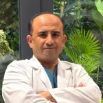
Ophthalmologist and eye surgeon. Graduate of Erciyes University Faculty of Medicine; completed his residency at Ankara Atatürk Training and Research Hospital. With 21 years of experience in cataract and refractive surgery, strabismus, glaucoma, retina and oculoplastic surgery, he sees patients at HG Hospital in Kahramanmaraş, Türkiye. Biography and training
Dr. Nejmi Öztürk
Ophthalmologist and eye surgeon
HG Hospital, Üngüt Mevkii, Prof. Dr. Necmettin Erbakan Blv. No:209, Onikişubat / Kahramanmaraş
+90 506 377 47 68WhatsAppAppointment formHospital switchboard (within Türkiye): 444 46 46 · info@nejmiozturk.com
- Private patients and private health insurance are welcome; patients with SGK coverage may use it (conditions are confirmed by the hospital)
- Same-day examination and test planning for patients travelling from other districts or from abroad
- Getting here and patient information
- Patients from Elbistan and the region