Eye Diseases and Treatments
By Dr. Nejmi Öztürk, ophthalmologist and eye surgeon, at HG Hospital in Kahramanmaraş, Türkiye — eye examinations for adults and children, medical and surgical treatments
- Short summaries of glaucoma, eyelid conditions (blepharoplasty and ptosis), uveitis, strabismus and lazy eye, dry eye, blocked tear duct, pterygium, blepharitis and corneal diseases
- What a general eye examination covers, when children should have their first examination, and glaucoma screening over the age of 40
- Which symptoms are emergencies; how an examination is arranged at HG Hospital in Kahramanmaraş
What eye diseases are there, and how are they treated?
Eye diseases can affect every layer of the eye, from the eyelids to the optic nerve; most can be treated if caught early. This page summarizes nine common topics apart from cataract, laser and retina; the details are on each topic’s own page. For cataract, see cataract surgery; for the retina, see retinal diseases.
Glaucoma (eye pressure)
Glaucoma is silent damage to the optic nerve, most often linked to high pressure inside the eye. Its most common form, open-angle glaucoma, causes no symptoms; loss begins in peripheral vision and cannot be reversed once it is noticed. Pressure inside the eye is usually between 10 and 21 mmHg, but nerve damage can also develop at normal values. For diagnosis, eye pressure measurement, corneal thickness, examination of the drainage angle, OCT analysis of the nerve fibers and a visual field test are assessed together. Treatment starts with drops; when needed, SLT laser, iridotomy or surgery (trabeculectomy, tube, MIGS) is used. Regular screening is recommended for everyone over 40. Details: Glaucoma (eye pressure).
Eyelids: blepharoplasty and ptosis
Ptosis (drooping eyelid) is when the upper eyelid drops far enough to partly cover the pupil; it can be present from birth or develop with age as the eyelid muscle loosens. In children it can lead to lazy eye, in adults to a narrowed field of vision and a habit of tilting the head back to see. Blepharoplasty is the removal of excess skin and fat from the eyelid; it is functional when the sagging restricts vision and cosmetic in other cases. Both operations are usually done under local anesthesia as outpatient procedures; swelling and bruising settle in 1–2 weeks. Ptosis that restricts vision may be covered by SGK under certain conditions; cosmetic blepharoplasty is not covered. Details: Blepharoplasty and ptosis.
Uveal diseases (uveitis)
Uveitis is inflammation of the vascular layer of the eye (the uvea). It presents with pain, redness, light sensitivity, blurring and floating spots; it can begin suddenly or gradually, in one or both eyes. It can be linked to rheumatic diseases, Behçet’s disease and some infections, so it is often followed together with the rheumatology department and blood tests are requested. Untreated uveitis can lead to cataract, glaucoma and macular edema. Treatment uses cortisone drops, drops that widen the pupil and, when needed, intravitreal injections and immune-modulating medicines. Because it tends to recur, regular follow-up is needed. Details: Uveal diseases.
Strabismus and lazy eye
Strabismus is when the eyes do not look at the same point; lazy eye (amblyopia) is when one eye fails to complete its visual development because of a glasses prescription, strabismus or a drooping eyelid. Lazy eye can be treated only in childhood, up to about the age of 8–9, so early diagnosis is decisive. Diagnosis involves measuring vision, a glasses examination with drops and examination of the eye movements. Treatment consists of glasses, covering the stronger eye (patching) and, when needed, strabismus surgery on the eye muscles. In children the operation is done under general anesthesia. In adults, strabismus surgery is done to correct double vision and the alignment of the eyes. Details: Strabismus and lazy eye.
Dry eye
Dry eye is when the surface of the eye is not kept moist enough because too little tear fluid is produced or it evaporates too quickly. Stinging, burning, a gritty feeling, redness, blurring at the end of the day and, paradoxically, watering in the wind are seen. Long screen use, air conditioning, age, menopause, certain medicines and inflammation of the eyelid margin increase the risk. For diagnosis, the amount of tear fluid and the tear break-up time are measured and the eyelid glands are assessed. Treatment starts with artificial tear drops, eyelid cleaning and warm compresses; when needed, anti-inflammatory drops and plugs for the tear ducts are used. It is a chronic condition that is kept under control with regular care. Details: Dry eye.
Blocked tear duct and DCR
A blocked tear duct is the closure of the channel that drains tears into the nose; it causes constant watering, discharge and repeated infections of the tear sac. In babies, a blockage present from birth is common and most open with massage by the age of one; if not, a short probing procedure is done. In adults with a permanent blockage, DCR (dacryocystorhinostomy) surgery creates a new passage between the tear sac and the nose. DCR can be done through a small incision on the skin or from inside the nose; it is performed under local or general anesthesia, and in most patients the watering largely resolves. Details: Blocked tear duct.
Pterygium (surfer’s eye)
A pterygium (“surfer’s eye”) is a triangular, vascular growth that extends from the white of the eye onto the cornea. Exposure to sun, wind and dust are the main causes; it is common in people who work outdoors and in the fields. It causes redness and stinging and, as it grows, astigmatism and reduced vision; at the examination it is distinguished from a pinguecula, a more harmless growth with a similar appearance. A small pterygium without symptoms is monitored with sunglasses and artificial tears. A pterygium approaching the visual axis or one that is constantly inflamed is removed surgically under local anesthesia; to reduce recurrence, a graft of the patient’s own conjunctiva is placed over the area. Details: Pterygium (surfer’s eye).
Blepharitis (inflammation of the eyelid margin)
Blepharitis (“inflammation of the eyelid margin”) is chronic inflammation of the oil glands at the edge of the eyelid and the roots of the eyelashes. Its main symptoms are crusting in the morning, dandruff-like flakes at the base of the lashes, itching, redness and recurrent styes; it is often seen together with dry eye and rosacea of the skin. The diagnosis is made with a slit-lamp examination. The basis of treatment is home care such as daily eyelid cleaning and warm compresses; when needed, antibiotic or anti-inflammatory drops and ointments and oral medicines are added. It is chronic; flare-ups are reduced with regular care, but it is not expected to disappear completely. Contact lens wear is continued with breaks during flare-ups. Details: Blepharitis.
Corneal diseases
The cornea is the clear layer at the front of the eye; it can lose its clarity through diseases such as infection (keratitis), injury, keratoconus (bulging of the cornea into a cone shape) and inherited dystrophies. Pain, light sensitivity, redness and blurring are the main symptoms; contact lens wearers have a high risk of infection, and painful redness is assessed urgently. Diagnosis uses the slit-lamp examination, corneal topography and thickness measurement; if infection is suspected, a culture may be taken. Depending on the cause, treatment may be antibiotic or anti-inflammatory drops, special contact lenses, cross-linking (CXL) to halt progression in keratoconus and, in advanced cases, a corneal transplant. Details: Corneal diseases and keratoconus treatment.
Which symptoms are emergencies?
With the following symptoms, see an eye doctor or go to the emergency department the same day, without waiting for an appointment:
- Sudden loss of vision, or vision closing off like a curtain
- Severe, painful redness; if headache and nausea accompany it, it may be an acute glaucoma attack
- Sudden flashes of light and a sudden increase in floaters (retinal tear)
- A chemical splash in the eye: rinse with plenty of water for 15–20 minutes, then seek care
- A blow to the eye, a cut, a foreign body; sudden double vision
- A white reflection in the pupil of a baby or child
Who needs regular examinations, and which tests are not suitable for everyone?
Even without symptoms, everyone over 40, people with diabetes or high blood pressure, people with a family history of glaucoma or macular degeneration and those taking cortisone benefit from regular examinations. Some tests are not suitable for everyone: the drops that dilate the pupil are used with care in eyes with narrow angles; dye angiography may be postponed in kidney failure, severe allergy and pregnancy.
What does a general eye examination cover?
A general eye examination is a 30–60 minute process that assesses every layer of the eye, from visual acuity to the back of the eye: your history; measurement of vision and glasses prescription (refraction); eye pressure (tonometry); examination of the eyelids, cornea and lens with the slit lamp; eye alignment; examination of the retina and optic nerve after drops; and, if needed, OCT, visual field test, corneal topography and tear tests.
How is an eye examination done?
- Your history is taken; your glasses, your list of medicines and any previous reports are reviewed.
- Visual acuity is measured and the glasses prescription is determined.
- Eye pressure is measured with an air-puff device or, after anesthetic drops, with a contact method; you feel a light puff or a brief touch.
- The front part of the eye is examined with the slit lamp; if needed, drops that dilate the pupil are instilled and there is a 20–30 minute wait.
- The back of the eye is examined; any further tests needed are done the same day.
- The results are explained; the treatment and the date of the next check are given to you in writing.
No anesthesia is needed for the examination. The dilated pupil blurs near vision for 4–6 hours; you should not drive during this time.
When should children have an eye examination?
Children should have their first eye examination at 6–12 months of age, even without symptoms; the second at 3–4 years (pre-school screening), the third before starting school, and then every 1–2 years. In premature babies, screening for retinopathy of prematurity (ROP) must be planned on time in the weeks after birth. In pre-school screening, vision is measured with picture tests; if lazy eye is caught at this age, the success of treatment is high.
A wandering eye, tilting the head to one side to look, squinting the eyes, sitting very close to the television and discomfort in light call for an examination regardless of age. In children the glasses prescription is measured with drops that relax the focusing muscle; their effect can last 1–3 days.
Why is glaucoma screening needed over the age of 40?
Because glaucoma progresses without symptoms and lost vision does not come back, eye pressure measurement and examination of the optic nerve are recommended every 2–4 years after the age of 40 and every 1–2 years after 60. People with a family history of glaucoma and those taking cortisone are screened earlier and more often. Cataract and macular changes are assessed at the same examination.
What are the risks and limitations?
- The drops that dilate the pupil cause blurring and light sensitivity for hours; rarely, in eyes with narrow angles, they can cause a rise in pressure.
- A single examination does not rule out every disease; glaucoma and macular degeneration develop over years and need regular follow-up.
- The risks of each treatment are described on its own page; surgical results vary from person to person.
- Evisceration, Enucleation and the Ocular Prosthesis
Which symptom suggests which disease?
| Symptom | Possible cause | Urgency |
|---|---|---|
| Silent loss of peripheral vision | Glaucoma | Planned, but should not be delayed |
| Painful redness, light sensitivity | Uveitis, keratitis | Urgent |
| A wandering eye in a child | Strabismus, lazy eye | Early examination |
| Stinging, burning, gritty feeling | Dry eye, blepharitis | Planned |
| Constant watering, discharge | Blocked tear duct | Planned; urgent if the tear sac is infected |
Eye examination and treatment in Kahramanmaraş
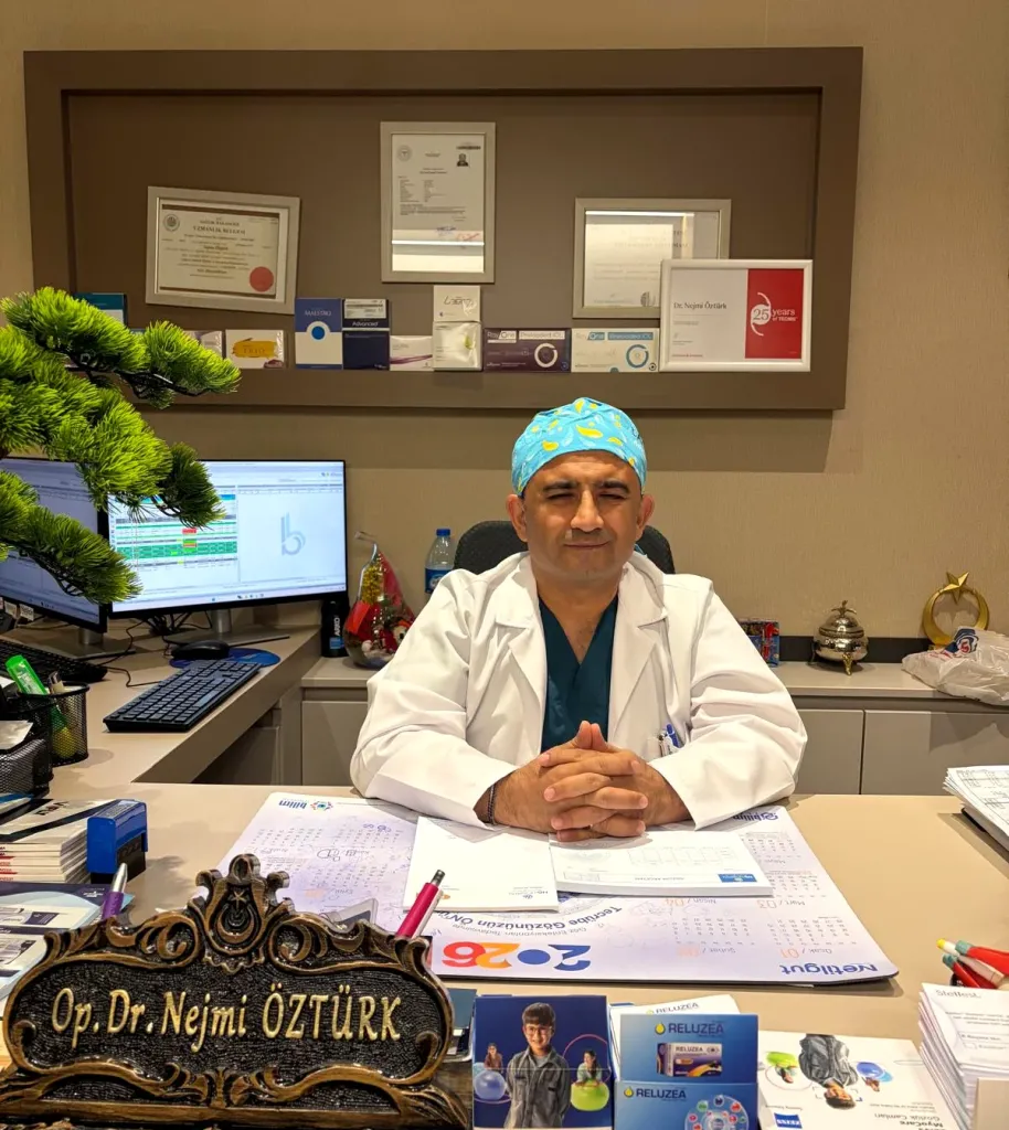
At HG Hospital in Kahramanmaraş, Türkiye, eye examinations for adults and children and the medical and surgical treatment of the diseases on this page are carried out by Dr. Nejmi Öztürk.
HG Hospital is a private hospital contracted with the Turkish social security institution (SGK); patients with valid SGK coverage may use it, although a private-hospital co-payment may apply. Eye examinations and most of these treatments are within SGK coverage; cosmetic blepharoplasty is not. Patients without SGK coverage, such as international patients and visitors from abroad, pay privately or through their private or travel health insurance; the hospital checks coverage before treatment. Fee information is given by the hospital after the examination, and insurance coverage is clarified at the consultation.
Dr. Öztürk sees patients only at HG Hospital in Kahramanmaraş. For those coming from Elbistan, Afşin, Göksun, Ekinözü and Nurhak, the examination and tests are grouped into the same day. For details, see the patients from Elbistan and the surrounding area page.
Dr. Nejmi Öztürk sees patients at HG Hospital in Kahramanmaraş, Türkiye. For patients travelling from other districts or from abroad, examination, tests and treatment planning can be arranged for the same day.
Hospital switchboard (within Türkiye): 444 46 46 · E-mail: info@nejmiozturk.com · HG Hospital, Üngüt Mevkii, Prof. Dr. Necmettin Erbakan Blv. No:209, Onikişubat / Kahramanmaraş, Türkiye
Frequently asked questions
How long does an eye examination take?
A general eye examination takes 30–60 minutes; if drops that dilate the pupil are used, a 20–30 minute wait is added. Further tests are done the same day.
When should my child have a first eye examination?
An examination is recommended at 6–12 months even without symptoms, then at 3–4 years and before starting school. If there is a wandering eye, squinting or discomfort in light, do not wait for these ages.
What should I bring to the examination?
Bring your glasses, your list of medicines, any previous eye reports and, if you have diabetes, your latest HbA1c result. Do not drive, because your pupils may be dilated.
Are dry eye and blepharitis the same disease?
They are different diseases but are often seen together; blepharitis disrupts the oily layer of the tear film and leads to dryness. Eyelid cleaning and warm compresses are part of the treatment of both.
Is eyelid surgery covered by SGK?
Ptosis surgery that restricts the field of vision may be within SGK coverage under certain conditions; cosmetic blepharoplasty is not covered by SGK or by most health insurance policies. Coverage is clarified at the consultation.
Does strabismus go away as a child grows?
In babies, temporary deviations in the first months can resolve by 4–6 months; a deviation that persists after that needs an examination. Untreated strabismus can lead to lazy eye, and lazy eye can be treated only in childhood.
How much does an examination cost?
Fee information is given by the hospital after the examination; insurance coverage is clarified at the consultation. A private-hospital co-payment may apply for SGK-insured patients; patients without SGK coverage pay privately or through their private or travel health insurance.
I am coming from Elbistan; will the examination and tests be finished the same day?
Yes; the examination and tests are planned for the same day. Because the drive takes about 2 hours, you are advised to book a morning appointment and to bear in mind that you will not be able to drive back because of the drops.
Sources
- Turkish Ophthalmological Society — todnet.org
- American Academy of Ophthalmology (AAO) — aao.org
- National Eye Institute (NEI) — nei.nih.gov
Related pages
This page is for information only; diagnosis and treatment decisions are made after an examination. The results of any surgical or interventional procedure may vary from person to person. You are advised to obtain detailed advice from your physician before any procedure.
Ophthalmologist and eye surgeon. Graduate of Erciyes University Faculty of Medicine; completed his residency at Ankara Atatürk Training and Research Hospital. With 21 years of experience in cataract and refractive surgery, strabismus, glaucoma, retina and oculoplastic surgery, he sees patients at HG Hospital in Kahramanmaraş, Türkiye. Biography and training
Dr. Nejmi Öztürk
Ophthalmologist and eye surgeon
HG Hospital, Üngüt Mevkii, Prof. Dr. Necmettin Erbakan Blv. No:209, Onikişubat / Kahramanmaraş
+90 506 377 47 68WhatsAppAppointment formHospital switchboard (within Türkiye): 444 46 46 · info@nejmiozturk.com
- Private patients and private health insurance are welcome; patients with SGK coverage may use it (conditions are confirmed by the hospital)
- Same-day examination and test planning for patients travelling from other districts or from abroad
- Getting here and patient information
- Patients from Elbistan and the region