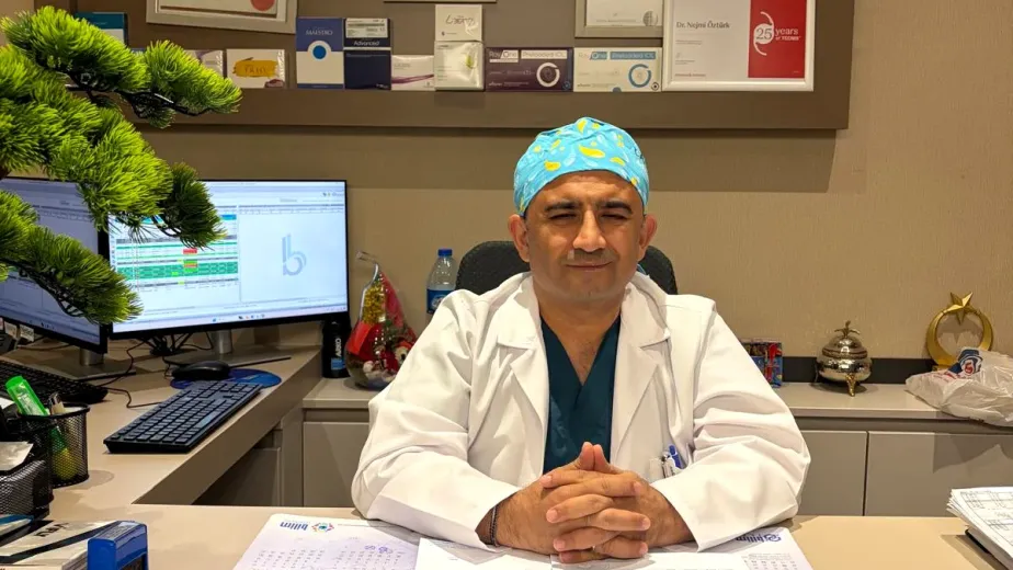
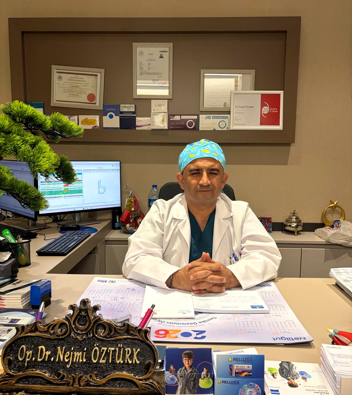
At Private HG Hospital in Kahramanmaraş, Türkiye, by Dr. Nejmi Öztürk, ophthalmologist — monitoring eye health from infancy to school age
- Timing of examinations — at which ages check-ups are recommended, from the newborn period to preschool
- Retinopathy of prematurity (ROP) — who is screened and why timing is decisive
- Warning signs — findings a family can notice at home
- Common eye problems in children
- School age and myopia — time outdoors, near work and screen habits
- What happens at a child’s eye examination
Vision is not an ability that arrives ready-made at birth; the visual centers of the brain mature in the early years of life, through the clear images that come from the eyes. For this reason, children’s eye health is less a matter of glasses prescriptions than of monitoring development: many problems noticed at a young age can be treated, whereas the same problem found after visual development is complete may allow limited gains.
Children often do not describe vision problems; a child who sees well with one eye does not complain even when the other eye sees poorly, because they are not struggling. This is why check-ups planned by age matter.
When should a baby’s eyes be examined?
The approach in children’s eye health is not to wait for symptoms but to check the eyes and the development of vision at set ages. The recommended steps are as follows:
- In the newborn period: A red reflex test should be done. In a darkened room a light is held to the eye, and the examiner checks whether an orange-red glow comes back from the back of the eye. If no glow is returned, if it looks dull, or if there is a clear difference between the two eyes, it can be a sign of congenital cataract, a tumor inside the eye or another condition affecting the back of the eye.
- Between 6 and 12 months: It is assessed whether the eyes move together, whether there is any turning, and the condition of the back of the eye (the fundus); the examiner observes whether the baby follows light and faces.
- Around age 3: Because the child can now be tested with shape and picture cards, the vision level of each eye can be measured separately; refractive errors (prescriptions) that need glasses, and lazy eye, can be caught at this stage.
- Preschool (ages 4–5): Visual acuity, refractive error and eye movements are re-evaluated. This check can prevent school difficulties caused by not being able to see the board.
- School age: Even without complaints, check-ups continue at intervals; refractive errors can change during growth.
If there is a family history of high prescriptions, lazy eye, strabismus (squint), congenital cataract or childhood glaucoma, these intervals are not waited for; the child should be evaluated earlier and more often. Timing may not be the same for every child.
Retinopathy of prematurity (ROP) screening in premature babies
Retinopathy of prematurity (ROP) is related to the blood vessels of the retina, the light-sensing layer at the back of the eye, not having finished developing in babies born before term. The retinal vessels mature in the last weeks of pregnancy; preterm birth interrupts this process and in some babies the vessels grow irregularly. The condition mostly regresses by itself, while in some babies it needs treatment.
ROP has no sign that a family can notice from the outside; the baby is not fussy, and no redness or discharge appears in the eye. The diagnosis is made solely by planned examination of babies in the risk group.
Who is screened?
- National guidelines cover all babies below certain thresholds of birth weight and gestational age; in Türkiye it is common for babies with a birth weight of 1500 grams or less, or a gestational age of 34 weeks or less, to fall into this group.
- Even if born above these thresholds, babies who received oxygen or breathing support for a long time, had a severe infection or had a complicated course in intensive care are screened on the referral of the neonatologist (the newborn specialist).
- Multiple pregnancy and growth restriction are also taken into account.
Why is timing decisive?
In ROP the course changes from week to week, and the stage that needs treatment has to be caught within a limited window of time. Examination therefore starts while the baby is still in intensive care: the general approach is an assessment at about 4 weeks after birth or around the calculated 31st postmenstrual week (gestational age plus the weeks since birth). The frequency of later check-ups can range from one to three weeks depending on the retinal finding; missing check-ups can mean the treatment window closes.
The examination is done after the pupil has been dilated with drops, using a light source and a special lens to inspect the retina including its outer edges; imaging can also be used so that the finding can be compared over time. Because the eyelids are gently held open, the baby may be unsettled during the short examination; this is expected. If treatment is needed, the method and timing are set according to the retinal finding and the baby’s general condition; results may vary from baby to baby.
The video below is a short recording made during an examination.
When screening is completed, follow-up does not end: children born early are more likely to have refractive errors, lazy eye and strabismus at older ages, and regular eye check-ups are recommended for them.
Warning signs a family can notice
If any of the following is seen, do not wait for the scheduled check-up; the child should be evaluated by an eye doctor.
- Eye turning (strabismus): One of the eyes turning in, out, up or down. Short-lived turning can be seen in the early months; if the turning is constant, obvious or continues after 4 months, it should be evaluated.
- Whiteness in the eye (leukocoria): The pupil looking white, grayish or cloudy; the red reflection of one eye missing in photographs. It should be evaluated without delay.
- Excessive sensitivity to light: Squeezing the eyes shut in bright light, constantly looking away, becoming fussy.
- Constant tearing or discharge: Tearing that continues even without crying, eyelids stuck together in the morning.
- Tilting the head to look: Always tilting the head in the same direction to look at the television, the board or a toy.
- Droopy eyelid: The lid partly covering the pupil; especially if there is a difference between the two eyes.
- Getting close and squinting: Moving too close to the screen, squinting when looking into the distance, holding a book very close to the eyes.
- Involuntary shaking of the eyes (nystagmus) and a marked reaction when one eye is covered: Being upset when one eye is covered but not reacting when the other is covered can be a sign of a difference in vision between the two eyes.
Each of these signs can arise from more than one cause; the distinction is made by examination.
Common eye problems in children
- Refractive errors: Myopia (nearsightedness — blurred distance vision), hyperopia (farsightedness — straining up close, eye fatigue) and astigmatism (blurring at every distance, headache). Glasses are given not just to sharpen the image but also to support visual development.
- Strabismus (squint): The eyes not looking parallel to each other. The cause may be a refractive error or may relate to how the eye muscles work; treatment may involve glasses, patching and, in suitable cases, surgery.
- Lazy eye (amblyopia): When a clear image does not reach the brain from one eye, the visual development of that eye lags behind; the cause may be a large difference in prescription between the eyes, strabismus or a condition that obstructs vision. The response to treatment is better when it is noticed early.
- Blocked tear duct: One of the common causes of constant tearing and discharge in babies. Most cases improve on their own in the early period with massage and hygiene measures; an intervention is considered in cases that do not resolve.
- Conjunctivitis (pink eye): Comes with redness, tearing, discharge and a stinging feeling; the cause can be a virus, bacteria or allergy, and treatment varies with the cause. Because it can be contagious, hand hygiene is important.
- Stye (hordeolum) and chalazion: Swellings that form when the oil glands at the eyelid margin become blocked or inflamed. Warm compresses and lid cleaning are the starting steps; lesions that do not go away need a doctor’s assessment.
- Congenital cataract: The lens of the eye being cloudy from birth; it is noticed by whiteness in the pupil or by the red reflex not being obtained, and because it directly affects visual development it should be evaluated in good time.
- Congenital glaucoma: A rare condition in which the pressure inside the eye rises. Excessive sensitivity to light, tearing and an eye that looks larger than usual are its typical signs; it should be evaluated without delay.
The short video below shares footage recorded during an examination.
School age and the rise in myopia
Studies in different countries have shown that myopia is more common in children and starts at younger ages. Myopia that begins in childhood tends to progress through growth; monitoring its progression is therefore as important as correcting the prescription with glasses.
Heredity has a clear share in the development of myopia: if one or both parents are myopic, the likelihood in the child rises. Beyond heredity, environmental habits have also been shown to play a role:
- Time spent outdoors: More time in daylight has been linked to a lower chance of myopia starting; in studies, an average of two hours a day stands out. This does not mean it reverses an existing prescription; the effect is more about delaying the onset.
- Uninterrupted near work: Focusing up close for a long time without a break increases eye fatigue; the common recommendation is to take a short break about every 20 minutes and look into the distance. Holding a book or screen too close and inadequate lighting also increase strain.
- Screen habits: Screen time matters, but so does the activity the screen replaces. Small screens are held closer, so screens that can be watched from a distance may be preferred to phones and tablets.
These recommendations cannot be presented as a way to prevent myopia; habits do not replace monitoring the prescription. In cases that progress, measures aimed at myopia control (low-dose atropine drops, or glasses and contact lens options designed for this purpose) may come up after the doctor’s assessment. Suitability is determined by the child’s age, the course of the prescription and the examination findings; the response varies from child to child.
What happens at a child’s eye examination?
A child’s eye examination is not a scaled-down adult examination; the method changes with age. Behavior-based methods are used in a baby who cannot speak, and letter and shape cards in a school-age child:
- History: Questions cover the pregnancy and birth, any history of premature birth and intensive care, a family history of glasses, lazy eye or strabismus, and the child’s behavior as observed at home and at school.
- Measuring the level of vision: The two eyes are assessed separately with age-appropriate cards; in young children, behavioral measures such as following a light and the reaction to covering one eye are used.
- Eye movements and alignment: Whether the eyes move together and the degree of any turning are measured with cover tests and prisms.
- Anterior segment (front of the eye) examination: The eyelids, lashes, conjunctiva, cornea, pupil and lens are examined under a biomicroscope (slit lamp).
- Refraction with drops (cycloplegic refraction): Because a child’s focusing muscle is very strong, the eye does not show its true prescription by itself; measured without drops, the prescription can come out lower than it really is. Drops that temporarily relax the focusing are therefore instilled, and the measurement is taken after about 30–45 minutes. The pupil gets larger, near vision stays blurred for a while and there is sensitivity to light; these are temporary.
- Fundus (back of the eye) examination: With the pupil dilated, the retina, the optic nerve and the blood vessels are assessed.
- Eye pressure: Where considered necessary, it is measured with methods suited to the child.
Preparation for the family is simple: the child should not be hungry, any previous glasses prescription and reports should be brought, and, since near vision stays blurred for a while after the examination, the plan for that day should be arranged accordingly.
A short recording made during an examination is in the video below.
Children’s eye examination in Kahramanmaraş
Eye examinations for babies and children, refraction with drops, assessment of eye turning and fundus examination are carried out at Private HG Hospital in Kahramanmaraş, Türkiye, by Dr. Öztürk, ophthalmologist. Because an examination with drops involves waiting, choosing an appointment time that suits the child’s routine makes the process easier.
For families coming from Elbistan, Afşin, Göksun, Ekinözü and Nurhak, the examination and tests are planned for the same day as far as possible, so that a second trip on the same road is not needed. It helps to mention this planning when booking; appointment and travel information is on the contact page. Diagnosis and treatment decisions are always made after an examination, and results may vary from child to child.
Frequently asked questions
My baby’s eyes cause no complaints; is an examination still needed?
Yes. Most vision problems in children cause no complaints; if one eye sees well, the child is not troubled and does not notice the problem. For this reason the red reflex test in the newborn period and check-ups at 6–12 months, at age 3 and in the preschool period are recommended regardless of complaints.
My baby’s eyes turn from time to time; is it right to wait?
Short-lived and changing turning can be seen in the early months. If the turning is constant, obvious, always in the same eye or continues after 4 months, an examination is needed. In some children the eye looks turned when it is not, because of a wide bridge of the nose; this is told apart by examination.
When is the eye examination done for my premature baby?
ROP screening usually begins while the baby is still in intensive care; the common approach is an assessment at about 4 weeks after birth or around the calculated 31st postmenstrual week. The frequency of check-ups is set according to the retinal finding; the planning is done jointly by the neonatologist and the eye doctor.
Does the ROP examination harm the baby?
The examination is done after the pupil has been dilated with drops, with the help of light and a lens, and it is short. Because the eyelids are gently held open, the baby may be unsettled; this is expected and passes after the examination.
Up to what age can lazy eye be treated?
The response to treatment is better in the years when visual development is continuing, which is why starting early matters. Treatment can also be tried after visual development is complete, but the gain may be limited, and it is assessed according to the examination findings.
Will the prescription increase if the child is given glasses?
Glasses do not increase the prescription; by letting the eye see clearly they support visual development and are part of treatment in the management of lazy eye and strabismus. It is normal for a child’s prescription to change with growth; this change comes from the development of the eye, not from the glasses.
Why is an examination with drops needed?
Because a child’s focusing muscle is very strong, the eye focuses by itself during measurement and does not show its true prescription; measured without drops, the prescription can come out lower than it really is. Drops that temporarily relax the focusing remove this error.
Can screen time permanently damage my child’s eyes?
Screen use is known to cause complaints such as eye fatigue, dryness and headache. In the development of myopia, heredity and time spent outdoors play a role together. During near work, looking into the distance about every 20 minutes, using the screen at an adequate distance and spending time outdoors are recommended.
I noticed whiteness in my child’s eye; what should I do?
Whiteness or cloudiness in the pupil (leukocoria), and the red reflection of one eye missing in photographs, are findings that need to be evaluated without delay; there is more than one possible cause, including congenital cataract.
How should we prepare for the child’s eye examination?
It helps if the child is not hungry or very tired and if any previous glasses prescription and reports are brought. If an examination with drops is to be done, you should expect to wait about 30–45 minutes and allow for near vision staying blurred for a while afterwards.
Sources
- Turkish Ophthalmological Society — todnet.org
- American Academy of Ophthalmology (AAO) — aao.org
- National Eye Institute (NEI) — nei.nih.gov
- World Health Organization — who.int
Related pages
- Strabismus (squint) and lazy eye
- Blocked tear duct
- Retinal diseases
- Eye doctor in Kahramanmaraş
- About Dr. Nejmi Öztürk
This article is for informational purposes; diagnosis and treatment decisions are made after an examination. The results of any treatment or interventional procedure may vary from person to person. You are advised to obtain your doctor’s opinion on decisions about your child’s eye health.
Ophthalmologist and eye surgeon. Graduate of Erciyes University Faculty of Medicine; completed his residency at Ankara Atatürk Training and Research Hospital. With 21 years of experience in cataract and refractive surgery, strabismus, glaucoma, retina and oculoplastic surgery, he sees patients at HG Hospital in Kahramanmaraş, Türkiye. Biography and training